Hostname: page-component-8448b6f56d-c4f8m Total loading time: 0 Render date: 2024-04-24T12:52:43.979Z Has data issue: false hasContentIssue false

Simultaneous Three-Dimensional Vascular and Tubular Imaging of Whole Mouse Kidneys With X-ray μCT

Published online by Cambridge University Press:  06 July 2020

Willy Kuo
Affiliation:
University of Zurich, Institute of Physiology, Winterthurerstrasse 190, 8057Zurich, Switzerland University of Zurich, National Centre of Competence in Research, Kidney. CH, Winterthurerstrasse 190, 8057Zurich, Switzerland University of Basel, Biomaterials Science Center, Department of Biomedical Engineering, Gewerbestrasse 14, 4123Allschwil, Switzerland
Ngoc An Le
Affiliation:
University of Zurich, Department of Chemistry, Winterthurerstrasse 190, 8057Zurich, Switzerland
Bernhard Spingler
Affiliation:
University of Zurich, Department of Chemistry, Winterthurerstrasse 190, 8057Zurich, Switzerland
Roland H. Wenger
Affiliation:
University of Zurich, Institute of Physiology, Winterthurerstrasse 190, 8057Zurich, Switzerland University of Zurich, National Centre of Competence in Research, Kidney. CH, Winterthurerstrasse 190, 8057Zurich, Switzerland
Anja Kipar
Affiliation:
University of Zurich, Laboratory for Animal Model Pathology (LAMP), Institute of Veterinary Pathology, Vetsuisse Faculty, Winterthurerstrasse 268, 8057Zurich, Switzerland
Udo Hetzel
Affiliation:
University of Zurich, Electron Microscopy Unit, Institute of Veterinary Pathology, Vetsuisse Faculty, Winterthurerstrasse 268, 8057Zurich, Switzerland
Georg Schulz
Affiliation:
University of Basel, Biomaterials Science Center, Department of Biomedical Engineering, Gewerbestrasse 14, 4123Allschwil, Switzerland
Bert Müller
Affiliation:
University of Basel, Biomaterials Science Center, Department of Biomedical Engineering, Gewerbestrasse 14, 4123Allschwil, Switzerland
Vartan Kurtcuoglu*
Affiliation:
University of Zurich, Institute of Physiology, Winterthurerstrasse 190, 8057Zurich, Switzerland University of Zurich, National Centre of Competence in Research, Kidney. CH, Winterthurerstrasse 190, 8057Zurich, Switzerland University of Zurich, Zurich Center for Integrative Human Physiology, 8057Zurich, Switzerland
*
*Author for correspondence: Vartan Kurtcuoglu, E-mail: vartan.kurtcuoglu@uzh.ch
Get access

Abstract

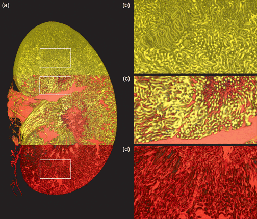
Concurrent three-dimensional imaging of the renal vascular and tubular systems on the whole-kidney scale with capillary level resolution is labor-intensive and technically difficult. Approaches based on vascular corrosion casting and X-ray micro computed tomography (μCT), for example, suffer from vascular filling artifacts and necessitate imaging with an additional modality to acquire tubules. In this work, we report on a new sample preparation, image acquisition, and quantification protocol for simultaneous vascular and tubular μCT imaging of whole, uncorroded mouse kidneys. The protocol consists of vascular perfusion with the water-soluble, aldehyde-fixable, polymeric X-ray contrast agent XlinCA, followed by laboratory-source μCT imaging and structural analysis using the freely available Fiji/ImageJ software. We achieved consistent filling of the entire capillary bed and staining of the tubules in the cortex and outer medulla. After imaging at isotropic voxel sizes of 3.3 and 4.4 μm, we segmented vascular and tubular systems and quantified luminal volumes, surface areas, diffusion distances, and vessel path lengths. This protocol permits the analysis of vascular and tubular parameters with higher reliability than vascular corrosion casting, less labor than serial sectioning and leaves tissue intact for subsequent histological examination with light and electron microscopy.

Type
Biological Applications
Copyright
Copyright © Microscopy Society of America 2020

Access options

Get access to the full version of this content by using one of the access options below. (Log in options will check for institutional or personal access. Content may require purchase if you do not have access.)

Footnotes

a

These authors contributed equally to the present study.

References

Adlam, D, De Bono, JP, Danson, EJ, Zhang, MH, Casadei, B, Paterson, DJ & Channon, KM (2011). Telemetric analysis of haemodynamic regulation during voluntary exercise training in mouse models. Exp Physiol 96(11), 11181128.CrossRefGoogle ScholarPubMed
Bankir, L, Figueres, L, Prot-Bertoye, C, Bouby, N, Crambert, G, Pratt, JH & Houillier, P (2020). Medullary and cortical thick ascending limb: Similarities and differences. Am J Physiol Renal Physiol 318(2), F422F442.Google ScholarPubMed
Beeuwkes, R III & Bonventre, JV (1975). Tubular organization and vascular-tubular relations in the dog kidney. Am J Physiol 229(3), 695713.CrossRefGoogle ScholarPubMed
Borgefors, G (1996). On digital distance transforms in three dimensions. Comput Vis Image Underst 64(3), 368376.CrossRefGoogle Scholar
Burgess, K, Jovanovic, S, Sudhir, R & Jovanovic, A (2019). Area under the curve analysis of blood pressure reveals increased spontaneous locomotor activity in SPAK knock-in mice: Relevance for hypotension induced by SPAK inhibition? Physiol Rep 7(3), e13997.CrossRefGoogle ScholarPubMed
Busse, M, Muller, M, Kimm, MA, Ferstl, S, Allner, S, Achterhold, K, Herzen, J & Pfeiffer, F (2018). Three-dimensional virtual histology enabled through cytoplasm-specific X-ray stain for microscopic and nanoscopic computed tomography. Proc Natl Acad Sci USA 115(10), 22932298.CrossRefGoogle ScholarPubMed
Chiefari, J, Chong, YK, Ercole, F, Krstina, J, Jeffery, J, Le, TPT, Mayadunne, RTA, Meijs, GF, Moad, CL, Moad, G, Rizzardo, E & Thang, SH (1998). Living free-radical polymerization by reversible addition−fragmentation chain transfer: The RAFT process. Macromolecules 31(16), 55595562.CrossRefGoogle Scholar
Chugh, BP, Lerch, JP, Yu, LX, Pienkowski, M, Harrison, RV, Henkelman, RM & Sled, JG (2009). Measurement of cerebral blood volume in mouse brain regions using micro-computed tomography. Neuroimage 47(4), 13121318.CrossRefGoogle ScholarPubMed
Czogalla, J, Schweda, F & Loffing, J (2016). The mouse isolated perfused kidney technique. J Vis Exp 117, 54712.Google Scholar
Ehling, J, Babickova, J, Gremse, F, Klinkhammer, BM, Baetke, S, Knuechel, R, Kiessling, F, Floege, J, Lammers, T & Boor, P (2016). Quantitative micro-computed tomography imaging of vascular dysfunction in progressive kidney diseases. J Am Soc Nephrol 27(2), 520532.CrossRefGoogle ScholarPubMed
Epah, J, Palfi, K, Dienst, FL, Malacarne, PF, Bremer, R, Salamon, M, Kumar, S, Jo, H, Schurmann, C & Brandes, RP (2018). 3D Imaging and quantitative analysis of vascular networks: A comparison of ultramicroscopy and micro-computed tomography. Theranostics 8(8), 21172133.CrossRefGoogle ScholarPubMed
Fan, L, Wang, S, He, X, Gonzalez-Fernandez, E, Lechene, C, Fan, F & Roman, RJ (2019). Visualization of the intrarenal distribution of capillary blood flow. Physiol Rep 7(8), e14065.CrossRefGoogle ScholarPubMed
Garcia-Sanz, A, Rodriguez-Barbero, A, Bentley, MD, Ritman, EL & Romero, JC (1998). Three-dimensional microcomputed tomography of renal vasculature in rats. Hypertension 31(1 Pt 2), 440444.CrossRefGoogle ScholarPubMed
Gardiner, BS, Smith, DW, O'Connor, PM & Evans, RG (2011). A mathematical model of diffusional shunting of oxygen from arteries to veins in the kidney. Am J Physiol Renal Physiol 300(6), F1339F1352.CrossRefGoogle ScholarPubMed
Ghanavati, S, Yu, LX, Lerch, JP & Sled, JG (2014). A perfusion procedure for imaging of the mouse cerebral vasculature by X-ray micro-CT. J Neurosci Methods 221, 7077.CrossRefGoogle ScholarPubMed
Grabherr, S, Djonov, V, Yen, K, Thali, MJ & Dirnhofer, R (2007). Postmortem angiography: Review of former and current methods. AJR Am J Roentgenol 188(3), 832838.Google ScholarPubMed
Grabherr, S, Hess, A, Karolczak, M, Thali, MJ, Friess, SD, Kalender, WA, Dirnhofer, R & Djonov, V (2008). Angiofil-mediated visualization of the vascular system by microcomputed tomography: A feasibility study. Microsc Res Technol 71(7), 551556.CrossRefGoogle ScholarPubMed
Haenssgen, K, Makanya, AN & Djonov, V (2014). Casting materials and their application in research and teaching. Microsc Microanal 20(2), 493513.CrossRefGoogle ScholarPubMed
Hall, AM, Rhodes, GJ, Sandoval, RM, Corridon, PR & Molitoris, BA (2013). In vivo multiphoton imaging of mitochondrial structure and function during acute kidney injury. Kidney Int 83(1), 7283.CrossRefGoogle ScholarPubMed
Hlushchuk, R, Zubler, C, Barre, S, Correa Shokiche, C, Schaad, L, Rothlisberger, R, Wnuk, M, Daniel, C, Khoma, O, Tschanz, SA, Reyes, M & Djonov, V (2018). Cutting-edge microangio-CT: New dimensions in vascular imaging and kidney morphometry. Am J Physiol Renal Physiol 314(3), F493F499.CrossRefGoogle ScholarPubMed
Hossler, FE & Douglas, JE (2001). Vascular corrosion casting: Review of advantages and limitations in the application of some simple quantitative methods. Microsc Microanal 7(3), 253264.CrossRefGoogle ScholarPubMed
Hossler, FE, Lametschwandtner, A, Kao, R & Finsterbusch, F (2013). Microvascular architecture of mouse urinary bladder described with vascular corrosion casting, light microscopy, SEM, and TEM. Microsc Microanal 19(6), 14281435.CrossRefGoogle ScholarPubMed
Hyde, DM, Tyler, NK & Plopper, CG (2007). Morphometry of the respiratory tract: Avoiding the sampling, size, orientation, and reference traps. Toxicol Pathol 35(1), 4148.CrossRefGoogle ScholarPubMed
Jin, E & Lu, ZR (2014). Biodegradable iodinated polydisulfides as contrast agents for CT angiography. Biomaterials 35(22), 58225829.Google ScholarPubMed
Klingberg, A, Hasenberg, A, Ludwig-Portugall, I, Medyukhina, A, Mann, L, Brenzel, A, Engel, DR, Figge, MT, Kurts, C & Gunzer, M (2017). Fully automated evaluation of total glomerular number and capillary tuft size in nephritic kidneys using lightsheet microscopy. J Am Soc Nephrol 28(2), 452459.CrossRefGoogle ScholarPubMed
Kolesova, H, Capek, M, Radochova, B, Janacek, J & Sedmera, D (2016). Comparison of different tissue clearing methods and 3D imaging techniques for visualization of GFP-expressing mouse embryos and embryonic hearts. Histochem Cell Biol 146(2), 141152.CrossRefGoogle ScholarPubMed
Kriz, W (1981). Structural organization of the renal medulla: Comparative and functional aspects. Am J Physiol 241(1), R3R16.Google ScholarPubMed
Krucker, T, Lang, A & Meyer, EP (2006). New polyurethane-based material for vascular corrosion casting with improved physical and imaging characteristics. Microsc Res Technol 69(2), 138147.CrossRefGoogle ScholarPubMed
Kuo, W & Kurtcuoglu, V (2017). Renal arteriovenous oxygen shunting. Curr Opin Nephrol Hypertens 26(4), 290295.CrossRefGoogle ScholarPubMed
Lai, JT, Filla, D & Shea, R (2002). Functional polymers from novel carboxyl-terminated trithiocarbonates as highly efficient RAFT agents. Macromolecules 35(18), 67546756.CrossRefGoogle Scholar
Lantuejoul, C & Beucher, S (1981). On the use of the geodesic metric in image-analysis. J Microsc 121, 3949.CrossRefGoogle Scholar
Le, NA, Kuo, W, Müller, B, Kurtcuoglu, V & Spingler, B (2020). Crosslinkable polymeric contrast agent for high-resolution X-ray imaging of the vascular system. Chem Commun 56(44), 58855888.Google ScholarPubMed
Legland, D, Arganda-Carreras, I & Andrey, P (2016). MorphoLibJ: Integrated library and plugins for mathematical morphology with ImageJ. Bioinformatics 32(22), 35323534.Google ScholarPubMed
Lusic, H & Grinstaff, MW (2013). X-ray-computed tomography contrast agents. Chem Rev 113(3), 16411666.Google ScholarPubMed
Ngo, JP, Kar, S, Kett, MM, Gardiner, BS, Pearson, JT, Smith, DW, Ludbrook, J, Bertram, JF & Evans, RG (2014). Vascular geometry and oxygen diffusion in the vicinity of artery–vein pairs in the kidney. Am J Physiol Renal Physiol 307(10), F1111F1122.CrossRefGoogle ScholarPubMed
Nordsletten, DA, Blackett, S, Bentley, MD, Ritman, EL & Smith, NP (2006). Structural morphology of renal vasculature. Am J Physiol Heart Circ Physiol 291(1), H296H309.CrossRefGoogle ScholarPubMed
Olgac, U & Kurtcuoglu, V (2015). Renal oxygenation: Preglomerular vasculature is an unlikely contributor to renal oxygen shunting. Am J Physiol Renal Physiol 308(7), F671F688.CrossRefGoogle ScholarPubMed
Olgac, U & Kurtcuoglu, V (2016). The Bohr effect is not a likely promoter of renal preglomerular oxygen shunting. Front Physiol 7, 482.CrossRefGoogle Scholar
Ollion, J, Cochennec, J, Loll, F, Escude, C & Boudier, T (2013). TANGO: A generic tool for high-throughput 3D image analysis for studying nuclear organization. Bioinformatics 29(14), 18401841.CrossRefGoogle ScholarPubMed
Ortiz, MC, Garcia-Sanz, A, Bentley, MD, Fortepiani, LA, Garcia-Estan, J, Ritman, EL, Romero, JC & Juncos, LA (2000). Microcomputed tomography of kidneys following chronic bile duct ligation. Kidney Int 58(4), 16321640.CrossRefGoogle ScholarPubMed
Pallone, TL, Zhang, Z & Rhinehart, K (2003). Physiology of the renal medullary microcirculation. Am J Physiol Renal Physiol 284(2), F253F266.CrossRefGoogle ScholarPubMed
Perrien, DS, Saleh, MA, Takahashi, K, Madhur, MS, Harrison, DG, Harris, RC & Takahashi, T (2016). Novel methods for microCT-based analyses of vasculature in the renal cortex reveal a loss of perfusable arterioles and glomeruli in eNOS−/− mice. BMC Nephrol 17, 24.CrossRefGoogle ScholarPubMed
Prommer, HU, Maurer, J, von Websky, K, Freise, C, Sommer, K, Nasser, H, Samapati, R, Reglin, B, Guimaraes, P, Pries, AR & Querfeld, U (2018). Chronic kidney disease induces a systemic microangiopathy, tissue hypoxia and dysfunctional angiogenesis. Sci Rep 8(1), 5317.CrossRefGoogle ScholarPubMed
Richardson, DS & Lichtman, JW (2015). Clarifying tissue clearing. Cell 162(2), 246257.CrossRefGoogle ScholarPubMed
Roberts, N, Magee, D, Song, Y, Brabazon, K, Shires, M, Crellin, D, Orsi, NM, Quirke, R, Quirke, P & Treanor, D (2012). Toward routine use of 3D histopathology as a research tool. Am J Pathol 180(5), 18351842.CrossRefGoogle ScholarPubMed
Schindelin, J, Arganda-Carreras, I, Frise, E, Kaynig, V, Longair, M, Pietzsch, T, Preibisch, S, Rueden, C, Saalfeld, S, Schmid, B, Tinevez, JY, White, DJ, Hartenstein, V, Eliceiri, K, Tomancak, P & Cardona, A (2012). Fiji: An open-source platform for biological-image analysis. Nat Methods 9(7), 676682.CrossRefGoogle ScholarPubMed
Schneider, CA, Rasband, WS & Eliceiri, KW (2012). NIH Image to ImageJ: 25 years of image analysis. Nat Methods 9(7), 671675.CrossRefGoogle ScholarPubMed
Stowell, RE (2009). Effect on tissue volume of various methods of fixation, dehydration, and embedding. Stain Technol 16(2), 6783.CrossRefGoogle Scholar
Tanaka, T & Nangaku, M (2013). Angiogenesis and hypoxia in the kidney. Nat Rev Nephrol 9(4), 211222.CrossRefGoogle ScholarPubMed
Tran, T, Sundaram, CP, Bahler, CD, Eble, JN, Grignon, DJ, Monn, MF, Simper, NB & Cheng, L (2015). Correcting the shrinkage effects of formalin fixation and tissue processing for renal tumors: Toward standardization of pathological reporting of tumor size. J Cancer 6(8), 759766.CrossRefGoogle ScholarPubMed
Vasquez, SX, Gao, F, Su, F, Grijalva, V, Pope, J, Martin, B, Stinstra, J, Masner, M, Shah, N, Weinstein, DM, Farias-Eisner, R & Reddy, ST (2011). Optimization of microCT imaging and blood vessel diameter quantitation of preclinical specimen vasculature with radiopaque polymer injection medium. PLoS One 6(4), e19099.CrossRefGoogle ScholarPubMed
Venkatachalam, MA, Weinberg, JM, Kriz, W & Bidani, AK (2015). Failed tubule recovery, AKI-CKD transition, and kidney disease progression. J Am Soc Nephrol 26(8), 17651776.CrossRefGoogle ScholarPubMed
Wagner, R, Van Loo, D, Hossler, F, Czymmek, K, Pauwels, E & Van Hoorebeke, L (2011). High-resolution imaging of kidney vascular corrosion casts with Nano-CT. Microsc Microanal 17(2), 215219.CrossRefGoogle ScholarPubMed
Wagner, RC, Czymmek, K & Hossler, FE (2006). Confocal microscopy, computer modeling, and quantification of glomerular vascular corrosion casts. Microsc Microanal 12(3), 262268.CrossRefGoogle ScholarPubMed
Wei, W, Popov, V, Walocha, JA, Wen, J & Bello-Reuss, E (2006). Evidence of angiogenesis and microvascular regression in autosomal-dominant polycystic kidney disease kidneys: A corrosion cast study. Kidney Int 70(7), 12611268.CrossRefGoogle ScholarPubMed
Yang, B, Treweek, JB, Kulkarni, RP, Deverman, BE, Chen, CK, Lubeck, E, Shah, S, Cai, L & Gradinaru, V (2014). Single-cell phenotyping within transparent intact tissue through whole-body clearing. Cell 158(4), 945958.CrossRefGoogle ScholarPubMed
Zhai, XY, Thomsen, JS, Birn, H, Kristoffersen, IB, Andreasen, A & Christensen, EI (2006). Three-dimensional reconstruction of the mouse nephron. J Am Soc Nephrol 17(1), 7788.CrossRefGoogle ScholarPubMed
Supplementary material: PDF

Kuo et al. supplementary material

Kuo et al. supplementary material

Download Kuo et al. supplementary material(PDF)
PDF 4.5 MB