Hostname: page-component-78c5997874-m6dg7 Total loading time: 0 Render date: 2024-11-18T01:15:44.071Z Has data issue: false hasContentIssue false

Quantitative non-destructive 3D Crystallographic Imaging of Microstructures using Laboratory X-ray Diffraction Contrast Tomography

Published online by Cambridge University Press:  10 August 2018

Hrishikesh Bale*
Affiliation:
Carl Zeiss Microscopy, Pleasanton, CA, USA.
Ron Kienan
Affiliation:
Dept. of Materials Science and Engineering, University of Michigan, Ann Arbor, MI, USA
Stephen T Kelly
Affiliation:
Carl Zeiss Microscopy, Pleasanton, CA, USA.
Nicolas Gueninchault
Affiliation:
Xnovo Technology ApS, K0ge, Denmark
Erik Lauridsen
Affiliation:
Xnovo Technology ApS, K0ge, Denmark
Ashwin Shahani
Affiliation:
Dept. of Materials Science and Engineering, University of Michigan, Ann Arbor, MI, USA
*
*Corresponding author, hrishikesh.bale@zeiss.com

Abstract

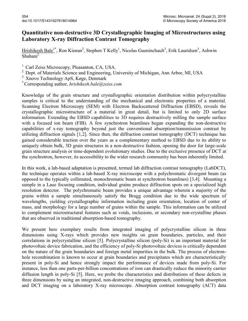
Image of the first page of this content. For PDF version, please use the ‘Save PDF’ preceeding this image.'
Type
Abstract
Copyright
© Microscopy Society of America 2018 

References

[1] Lienert, U, et al., Journal of the Minerals, Metals & Materials Society 63 2011) p. 70.Google Scholar
[2] Juul Jensen, D Poulsen, H.F. Materials Characterization 72 2012) p. 1.CrossRefGoogle Scholar
[3] Holzner, C, et al, Microscopy Today 24 2016) p. 34.Google Scholar
[4] McDonald, SA, et al, Scientific Reports 7 2017) p. 5251.Google Scholar
[5] Keinan, R, et al, Acta Materialia 148 2018) p. 255.Google Scholar