Hostname: page-component-8448b6f56d-sxzjt Total loading time: 0 Render date: 2024-04-19T17:45:57.998Z Has data issue: false hasContentIssue false

High-Energy Electron Scattering in Thick Samples Evaluated by Bright-Field Transmission Electron Microscopy, Energy-Filtering Transmission Electron Microscopy, and Electron Tomography

Published online by Cambridge University Press:  28 March 2022

Misa Hayashida*
Affiliation:
Nanotechnology Research Centre, National Research Council, Edmonton, AB T6G 2M9, Canada
Marek Malac
Affiliation:
Nanotechnology Research Centre, National Research Council, Edmonton, AB T6G 2M9, Canada Department of Physics, University of Alberta, Edmonton, AB T6G 2E1, Canada
*
*Corresponding author: Misa Hayashida, E-mail: misa.hayashida@nrc-cnrc.gc.ca
Get access

Abstract

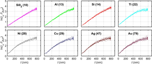
Energy-filtering transmission electron microscopy (TEM) and bright-field TEM can be used to extract local sample thickness $t$ and to generate two-dimensional sample thickness maps. Electron tomography can be used to accurately verify the local $t$. The relations of log-ratio of zero-loss filtered energy-filtering TEM beam intensity ($I_{{\rm ZLP}}$) and unfiltered beam intensity ($I_{\rm u}$) versus sample thickness $t$ were measured for five values of collection angle in a microscope equipped with an energy filter. Furthermore, log-ratio of the incident (primary) beam intensity ($I_{\rm p}$) and the transmitted beam $I_{{\rm tr}}$ versus $t$ in bright-field TEM was measured utilizing a camera before the energy filter. The measurements were performed on a multilayer sample containing eight materials and thickness $t$ up to 800 nm. Local thickness $t$ was verified by electron tomography. The following results are reported:

• The maximum thickness $t_{{\rm max}}$ yielding a linear relation of log-ratio, $\ln ( {I_{\rm u}}/{I_{{\rm ZLP}}})$ and $\ln ( {I_{\rm p}}/{I_{{\rm tr}}} )$, versus $t$.

• Inelastic mean free path ($\lambda _{{\rm in}}$) for five values of collection angle.

• Total mean free path ($\lambda _{{\rm total}}$) of electrons excluded by an angle-limiting aperture.

$\lambda _{{\rm in}}$ and $\lambda _{{\rm total}}$ are evaluated for the eight materials with atomic number from $\approx$10 to 79.

The results can be utilized as a guide for upper limit of $t$ evaluation in energy-filtering TEM and bright-field TEM and for optimizing electron tomography experiments.

Type
Software and Instrumentation
Copyright
Copyright © The Author(s), 2022. Published by Cambridge University Press on behalf of the Microscopy Society of America

Access options

Get access to the full version of this content by using one of the access options below. (Log in options will check for institutional or personal access. Content may require purchase if you do not have access.)

References

Cosslett, VE & Thomas, R (1964 a). Multiple scattering of 5–30 keV electrons in evaporated metal films: I. Total transmission and angular distribution. Br J Appl Phys 15, 883.CrossRefGoogle Scholar
Cosslett, VE & Thomas, RN (1964 b). Multiple scattering of 5–30 keV electrons in evaporated metal films II. Range-energy relations. Br J Appl Phys 15, 1283.CrossRefGoogle Scholar
De Backer, A, Van Aert, S, Nellist, PD & Jones, L (2021). Procedure for 3D atomic resolution reconstructions using atom-counting and a Bayesian genetic algorithm. Preprint. Available at arXiv:210505562.Google Scholar
de Jonge, N, Verch, A & Demers, H (2018). The influence of beam broadening on the spatial resolution of annular dark field scanning transmission electron microscopy. Microsc Microanal 24, 816.CrossRefGoogle ScholarPubMed
Drees, H, Müller, E, Dries, M & Gerthsen, D (2018). Electron-beam broadening in amorphous carbon films in low-energy scanning transmission electron microscopy. Ultramicroscopy 185, 6571.CrossRefGoogle ScholarPubMed
Egerton, R & Crozier, P (1989). Mass-thickness determination by Bethe-sum-rule normalization of the electron energy-loss spectrum. Ultramicroscopy 27, 918.Google Scholar
Egerton, R & Crozier, P (1997). The effect of lens aberrations on the spatial resolution of an energy-filtered TEM images. Micron 28, 117.CrossRefGoogle Scholar
Egerton, R, Li, P & Malac, M (2004). Radiation damage in the TEM and SEM. Micron 35, 399409.CrossRefGoogle ScholarPubMed
Egerton, R & Wong, K (1995). Some practical consequences of the Lorentzian angular distribution of inelastic scattering. Ultramicroscopy 59, 169180.CrossRefGoogle Scholar
Egerton, RF (2007). Limits to the spatial, energy and momentum resolution of electron energy-loss spectroscopy. Ultramicroscopy 107, 575586.CrossRefGoogle Scholar
Egerton, RF (2011). Electron Energy-Loss Spectroscopy in the Electron Microscope. Springer Science & Business Media.CrossRefGoogle Scholar
Everhart, TE & Hoff, PH (1971). Determination of kilovolt electron energy dissipation vs penetration distance in solid materials. J Appl Phys 42, 5837.CrossRefGoogle Scholar
Fitting, HJ (1974). Transmission, energy distribution and SE excitation of fast electrons in thin solid films. Phys Stat Solidi A 26, 525.CrossRefGoogle Scholar
Fujii, T, Malac, M, Kano, E, Hayashida, M, Yaguchi, T & Egerton, R (2018). Toward quantitative bright field TEM imaging of ultra thin samples. Microsc Microanal 24, 16121613.CrossRefGoogle Scholar
Gauvin, R & Rudinsky, S (2016). A universal equation for computing the beam broadening of incident electrons in thin films. Ultramicroscopy 167, 2130.CrossRefGoogle ScholarPubMed
Gouldsmit, S & Saunderson, J (1940). Multiple scattering of electrons. Phys Rev 57, 24.CrossRefGoogle Scholar
Grogger, W, Varela, M, Ristau, R, Schaffer, B, Hofer, F & Krishnan, KM (2005). Energy-filtering transmission electron microscopy on the nanometer length scale. J Electron Spectrosc 143, 139147.CrossRefGoogle Scholar
Hayashida, M, Cui, K, Homeniuk, D, Phengchat, R, Blackburn, AM & Malac, M (2019). Parameters affecting the accuracy of nanoparticle shape and size measurement in 3D. Micron 123, 102680.CrossRefGoogle ScholarPubMed
Hayashida, M & Malac, M (2016). Practical electron tomography guide: Recent progress and future opportunities. Micron 91, 4974.CrossRefGoogle ScholarPubMed
Hayashida, M, Malac, M, Bergen, M, Egerton, R & Li, P (2014 a). Accurate measurement of relative tilt and azimuth angles in electron tomography: A comparison of fiducial marker method with electron diffraction. Rev Sci Instrum 85, 083704.CrossRefGoogle ScholarPubMed
Hayashida, M, Malac, M, Bergen, M & Li, P (2014 b). Nano-dot markers for electron tomography formed by electron beam-induced deposition: Nanoparticle agglomerates application. Ultramicroscopy 144, 5057.CrossRefGoogle ScholarPubMed
Hayashida, M, Paraguay-Delgado, F, Ornelas, C, Herzing, A, Blackburn, AM, Haydon, B, Yaguchi, T, Wakui, A, Igarashi, K, Suzuki, Y, Motoki, S, Aoyama, Y, Konyuba, Y & Malac, M (2021). Nanoparticle size and 3d shape measurement by electron tomography: An inter-laboratory comparison. Micron 140, 102956.CrossRefGoogle ScholarPubMed
Hugenschmidt, M, Muller, E & Gersten, D (2019). Electron beam broadening in electron-transparent samples at low electron energies. J Microsc 274, 150157.CrossRefGoogle ScholarPubMed
Iakoubovskii, K, Mitsuishi, K, Nakayama, Y & Furuya, K (2008 a). Mean free path of inelastic electron scattering in elemental solids and oxides using transmission electron microscopy: Atomic number dependent oscillatory behavior. Phys Rev B 77, 104102.CrossRefGoogle Scholar
Iakoubovskii, K, Mitsuishi, K, Nakayama, Y & Furuya, K (2008 b). Thickness measurements with electron energy loss spectroscopy. Microsc Res Tech 71, 626631.CrossRefGoogle ScholarPubMed
Irmak, EA, Liu, P, Bals, S & Aert, SV (2021). 3D atomic structure of supported metallic nanoparticles estimated from 2D ADF STEM images: A combination of atom-counting and a local minima search algorithm. Small Methods 5, 2101150.CrossRefGoogle Scholar
Kanaya, K & Okayama, S (1972). Penetration and energy-loss theory of electrons in solid targets. J Phys D: Appl Phys 4, 43.CrossRefGoogle Scholar
Kato, M, Kawase, N, Kaneko, T, Toh, S, Matsumura, S & Jinnai, H (2008). Maximum diameter of the rod-shaped specimen for transmission electron microtomography without the missing wedge. Ultramicroscopy 108, 221229.CrossRefGoogle ScholarPubMed
Kohl, H & Reimer, L (2008). Transmission Electron Microscopy: Physics of Image Formation. Springer.Google Scholar
LeBeau, J, Findlay, S, Allen, L & Stemmer, S (2010). Standardless atom counting in scanning transmission electron microscopy. Nano Lett 10, 4054408.CrossRefGoogle ScholarPubMed
Lukiyanova, FA, Raub, EI & Sennov, RA (2009). Depth range of primary electrons, electron beam broadening, and spatial resolution in electron-beam studies. Bull Russ Acad Sci: Phys 73, 441449.CrossRefGoogle Scholar
Malac, M, Hettler, S, Hayashida, M, Kano, E, Egerton, R & Beleggia, M (2021). Phase plates in the transmission electron microscope: Operating principles and applications. Microscopy 70, 75115.CrossRefGoogle ScholarPubMed
Malac, M, Homeniuk, D, Hayashida, M, Fujii, T, Yaguchi, T & Egerton, R (2019). In-situ mass thickness calibrations using MWCNTs. Microsc Microanal 25, 792793.CrossRefGoogle Scholar
Malis, T, Cheng, S & Egerton, R (1988). EELS log-ratio technique for specimen-thickness measurement in the TEM. J Electron Microsc Tech 8, 193200.CrossRefGoogle ScholarPubMed
Matsukawa, T, Shimizu, R, Harada, K & Kato, T (1974). Investigation of kilovolt electron energy dissipation in solids. J Appl Phys 45, 733740.CrossRefGoogle Scholar
Ortega, E, Boothroyd, C & de Jonge, N (2021). The influence of chromatic aberration on the dose-limited spatial resolution of transmission electron microscopy. Ultramicroscopy 230, 113383.CrossRefGoogle ScholarPubMed
Pfaff, M, Muller, E, Klein, M, Colsmann, A, Lemmer, U, Krzyzanek, V, Reichelt, R & Gersten, D (2011). Low-energy electron scattering in carbon-based materials analyzed by scanning transmission electron microscopy and its application to sample thickness determination. Microscopy 243, 3139.CrossRefGoogle ScholarPubMed
Reimer, L & Sommer, K (1968). Messungen und berechnungen zum elektronenmikroskopischen streukontrast für 17 bis 1200 keV-elektronen. Z Naturforsch A 23, 15691582.CrossRefGoogle Scholar
Saghi, Z, Xu, X & Mobus, G (2008). Electron tomography of regularly shaped nanostructures under non-linear image acquisition. J Microsc 232, 186195.CrossRefGoogle ScholarPubMed
Sun, C, Müller, E, Meffert, M & Gerthsen, D (2018). On the progress of scanning transmission electron microscopy (STEM) imaging in a scanning electron microscope. Microsc Microanal 24, 99106.CrossRefGoogle Scholar
Thornton, JA (1974). Influence of apparatus geometry and deposition conditions on the structure and topography of thick sputtered coatings. J Vac Sci Technol 11, 666.CrossRefGoogle Scholar
Thornton, JA (1977). High rate thick film growth. Ann Rev Mater Sci 7, 239260.CrossRefGoogle Scholar
Wall, J & Hanfield, J (1986). Mass mapping with the scanning transmission electron microscope. Annu Rev Biophys Biophys Chem 15, 355376.CrossRefGoogle ScholarPubMed
Wittry, DB & Kyser, DF (1967). Measurement of diffusion lengths in direct-gap semiconductors by electron-beam excitation. J Appl Phys 38, 375.CrossRefGoogle Scholar
Yaguchi, T, Konno, M, Kamino, T & Watanabe, M (2008). Observation of three-dimensional elemental distributions of a Si device using a 360$^{\circ }$-tilt FIB and the cold field-emission STEM system. Ultramicroscopy 108, 16031615.CrossRefGoogle Scholar
Yamasaki, J, Mutoh, M, Ohta, S, Yuasa, S, Arai, S, Sasaki, K & Tanaka, N (2014). Analysis of nonlinear intensity attenuation in bright-field TEM images for correct 3D reconstruction of the density in micron-sized materials. Microscopy 63, 345355.CrossRefGoogle ScholarPubMed
Yamasaki, J, Ubata, Y & Yasuda, H (2019). Empirical determination of transmission attenuation curves in mass-thickness contrast TEM imaging. Ultramicroscopy 200, 2027.CrossRefGoogle ScholarPubMed
Yamashita, S, Kikkawa, J, Yanagisawa, K, Nagai, T, Ishizuka, K & Kimoto, K (2018). Atomic number dependence of $Z$ contrast in scanning transmission electron microscopy. Sci Rep 8, 12325.CrossRefGoogle ScholarPubMed
Yamashita, S, Koshiya, S, Ishizuka, K & Kimoto, K (2015 a). Quantitative annular dark-field imaging of single-layer graphene. Microscopy 64, 143150.CrossRefGoogle ScholarPubMed
Yamashita, S, Koshiya, S, Nagai, T, Kikkawa, J, Ishizuka, K & Kimoto, K (2015 b). Quantitative annular dark-field imaging of single-layer graphene-II: Atomic-resolution image contrast. Microscopy 64, 409418.CrossRefGoogle ScholarPubMed
Zhang, HR, Egerton, RF & Malac, M (2012). Local thickness measurement through scattering contrast and electron energy-loss spectroscopy. Micron 43, 815.CrossRefGoogle ScholarPubMed
Zhong, Z, Aveyard, R, Rieger, B, Bals, S, Palenstijn, WJ & Batenburg, KJ (2018). Automatic correction of nonlinear damping effects in HAADF–STEM tomography for nanomaterials of discrete compositions. Ultramicroscopy 184, 5765.CrossRefGoogle ScholarPubMed