Hostname: page-component-848d4c4894-pjpqr Total loading time: 0 Render date: 2024-06-26T04:56:49.953Z Has data issue: false hasContentIssue false

Autofluorescence For Rapid Visualization of Plant Anatomy Among Diverse Taxa

Published online by Cambridge University Press:  30 July 2021

Timothy Pegg
Affiliation:
Miami University, Oxford, Ohio, United States
Daniel Gladish
Affiliation:
Miami University, Oxford, Ohio, United States
Robert Baker
Affiliation:
Miami University, Oxford, Ohio, United States

Abstract

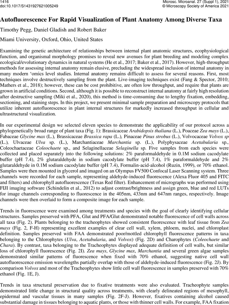
Image of the first page of this content. For PDF version, please use the ‘Save PDF’ preceeding this image.'
Type
To Fix or Not To Fix? A Question for Biological Samples
Copyright
Copyright © The Author(s), 2021. Published by Cambridge University Press on behalf of the Microscopy Society of America

References

Baker, R. L., Yarkhunova, Y., Vidal, K., Ewers, B. E. & Weinig, C. (2017). Polyploidy and the relationship between leaf structure and function: implications for correlated evolution of anatomy, morphology, and physiology in Brassica. BMC Plant Biology 17, 3. https://doi.org/10.1186/s12870-016-0957-3.CrossRefGoogle ScholarPubMed
Fang, Y. & Spector, D. L. (2010). Live Cell Imaging of Plants. Cold Spring Harbor Protocols 2010, pdb.top68. http://cshprotocols.cshlp.org/content/2010/2/pdb.top68.abstract.Google Scholar
He, W., Adachi, S., Sage, R. F., Ookawa, T. & Hirasawa, T. (2017). Leaf photosynthetic rate and mesophyll cell anatomy changes during ontogenesis in backcrossed indica × japonica rice inbred lines. Photosynthesis Research 134, 2738. https://doi.org/10.1007/s11120-017-0403-x.CrossRefGoogle ScholarPubMed
Li, Y. & Chen, L. (2014). Big biological data: challenges and opportunities. Genomics, proteomics & bioinformatics 12, 187189. https://pubmed.ncbi.nlm.nih.gov/25462151.Google ScholarPubMed
Mathers, A. W., Hepworth, C., Baillie, A. L., Sloan, J., Jones, H., Lundgren, M., Fleming, A. J., Mooney, S. J. & Sturrock, C. J. (2018). Investigating the microstructure of plant leaves in 3D with lab-based X-ray computed tomography. Plant Methods 14, 99. https://doi.org/10.1186/s13007-018-0367-7.CrossRefGoogle ScholarPubMed
Miki, Y., Saito, S., Niki, T. & Gladish, D. K. (2020). Three-dimensional digital image construction of metaxylem vessels in root tips of Zea mays subsp. mexicana from thin transverse sections. Applications in Plant Sciences 8, e11347. https://doi.org/10.1002/aps3.11347.Google ScholarPubMed
Ruzin, S. E. (1999). Plant microtechnique and microscopy. Oxford University Press New York.Google Scholar
Schindelin, J., Arganda-Carreras, I., Frise, E., Kaynig, V., Longair, M., Pietzsch, T., Preibisch, S., Rueden, C., Saalfeld, S., Schmid, B., Tinevez, J.-Y., White, D. J., Hartenstein, V., Eliceiri, K., Tomancak, P. & Cardona, A. (2012). Fiji: an open-source platform for biological-image analysis. Nature Methods 9, 676682. https://doi.org/10.1038/nmeth.2019.CrossRefGoogle ScholarPubMed