


Introduction
Species of Thelocarpon Nyl. are free living or lichenized fungi with minute perithecioid or apothecioid ascomata. Many species within the genus appear as pioneers, settling on bare or temporary substrata such as pebbles, clay or wood and typically disappear quickly again due to succession and subsequent competition or life-cycle completion (Kocourková-Horáková Reference Kocourková-Horáková1998). As a result, even though some species are widespread (e.g. Hafellner Reference Hafellner2017), most species are rarely collected. Species descriptions, especially in the early days, have resulted in numerous synonyms, most of which were resolved by Salisbury (Reference Salisbury1966).
The ascomata of Thelocarpon species typically contain flask-shaped multispored asci with colourless single-celled ascospores, although exceptions exist such as cylindrical asci (e.g. Thelocarpon lichenicola (Fuckel) Poelt & Hafellner) and 8-spored asci with 3-septate spores (Thelocarpon triseptatum Aptroot & M. Cáceres). Important characteristics that are used to distinguish species within Thelocarpon are the presence or absence of an algal sheath, periphyses and paraphyses. Some species develop verrucae with an algal sheath around the ascomata, but other species are only loosely associated with algae or not associated with algae at all. This variation in lichenization at times blurs the demarcation between free-living fungi and lichens within the genus. Filaments can be present in the ostiolar canal (periphyses), inside on the sides of the excipulum (periphysoids) and between the asci as interascal filaments (paraphyses) (Navarro-Rosinés et al. Reference Navarro-Rosinés, Roux and Bellemère1999), and their shape and presence are important characteristics that define the species.
Salisbury (Reference Salisbury1966) monographed the genus and recognized 13 Thelocarpon species, but many new species have been described since then. The type species of the genus, Thelocarpon laureri (Flot.) Nyl., has perithecioid ascomata that are enclosed within a verruca containing algae. Species with an exposed disc (e.g. Thelocarpon impressellum Nyl.) were transferred to the genus Ahlesia Fuckel (Salisbury Reference Salisbury1966), but are today again placed in Thelocarpon since the division of the genus based on disc exposure alone is considered arbitrary (Poelt & Hafellner Reference Poelt and Hafellner1975). However, the genus is probably heterogenous and several distinct subgroups can be distinguished (Salisbury Reference Salisbury1966; Navarro-Rosinés et al. Reference Navarro-Rosinés, Roux and Bellemère1999). Currently, DNA sequences from only a small number of species are available, and the phylogenetic position of the genus within the Pezizomycotina is still uncertain (Reeb et al. Reference Reeb, Lutzoni and Roux2004; Lumbsch et al. Reference Lumbsch, Zimmermann and Schmitt2009).
In the Netherlands, lichenologists have frequently collected and studied Thelocarpon species. For example, seven different species were reported by Aptroot & Sparrius (Reference Aptroot and Sparrius2000) in a single year. In recent years, Thelocarpon laureri and T. lichenicola have been collected in the Netherlands most often, both being recorded at around 20 localities since 2010. Species recorded in the Netherlands since then are Thelocarpon coccosporum Lettau, T. epibolum Nyl., T. impressellum, T. intermediellum Nyl., T. magnussonii G. Salisb., T. olivaceum B. de Lesd. and T. pallidum G. Salisb. In November 2021, an unknown species of Thelocarpon was found in a marl (soft calcareous rock) quarry in the Netherlands. It was different from other Thelocarpon species by having immersed brown perithecia with a green-yellowish ring around the ostiole, remarkably abundant and long periphyses and periphysoids, and wide, oblong and often somewhat asymmetrical ascospores. The same species was later found on marlstone at two different locations. The three collections consistently showed the same diagnostic characteristics, and hence the species is formally described here. Furthermore, since the monograph of Salisbury (Reference Salisbury1966), there has not been an up to date worldwide overview of Thelocarpon, therefore a key to all currently accepted Thelocarpon species is provided here.
Methods
Three specimens of the new Thelocarpon species were collected between 2021 and 2023 in the Netherlands. The collections were examined with a stereo microscope (Euromex SB 1903 P StereoBlue) and a compound microscope (Euromex iScope IS.1153-PLi). All measurements and observations were made on material mounted in water, except when stated otherwise. To test colour reactions, bleach (C), 10% KOH solution (K), para-phenylenediamine solution (P) and 1% or 10% Lugol's solution (I) were used. K/I represents the I reaction after application of a 10% KOH solution.
Taxonomy
Thelocarpon periphysatum Kolk, Timans, Boers & Sparrius sp. nov.
MycoBank No.: MB 848874
Differing from Thelocarpon immersum Fryday and T. opertum J. C. David & Coppins by the longer periphyses and periphysoids, up to 120 μm, and the oblong ascospores.
Type: The Netherlands, Limburg, Berg en Terblijt, Groeve Blom, on cyanobacterial crusts on marl in abandoned quarry, 50.8570°N, 5.7927°E, 21 November 2022, H. van der Kolk 3271 (L—holotype; hb. H. van der Kolk—isotype).
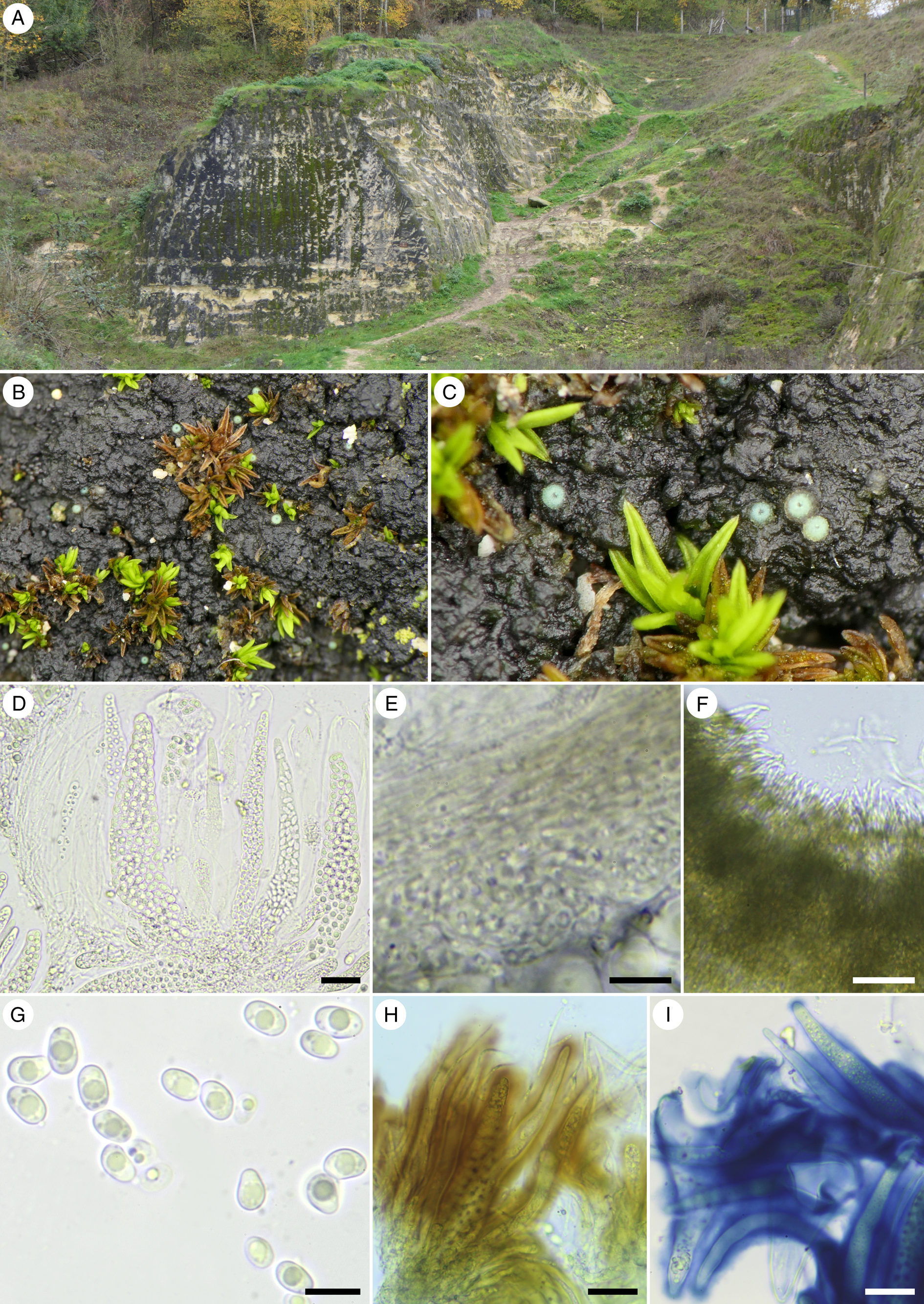
Figure 1. Thelocarpon periphysatum. A, habitat, marble walls in abandoned quarry. B & C, perithecia with distinct green-yellowish ring with bluish tinge immersed in crust of cyanobacteria. D, asci (note the long periphysoids on the left and the lack of true interascal filaments). E, excipulum. F, periphyses around the ostiole. G, ascospores. H, reaction of hymenial gel in I (Lugol's solution). I, reaction of hymenial gel in K/I. Scales: D–F, H & I = 20 μm; G = 10 μm. In colour online.

Figure 2. Thelocarpon periphysatum. A, schematic section of a perithecium; the periphysoids are more appressed to the exciple than appears in the drawing. B, ascospores. Scales: A = 100 μm; B = 10 μm. In colour online.
Thallus absent. Photobiont absent, but perithecia immersed in cyanobacterial crusts with Gloeocapsa-like and other species of cyanobacteria. All parts C‒, K‒ and P‒.
Ascomata perithecioid, globose, partially to completely immersed in cyanobacterial crusts with only the apices protruding, 200‒300 μm diam., without an exposed disc, opening through a small apical pore, light brown to brown, with a well-defined bright green-yellowish ring around the ostiole, with a bluish tinge in fresh material. Excipulum hyaline or light brown, 20‒35 μm thick. Periphyses and periphysoids abundant, arranged around the ostiole and on the sides, varying in length from 20 μm in the ostiolar canal to 120 μm further down the sides, then resembling paraphyses but not occurring between the asci, 1.0‒1.5 μm thick, simple. Hymenial gel hemiamyloid, I+ yellow (at low concentration) to I+ red (at high concentration), K/I+ blue. Paraphyses absent. Asci multispored (> 100 ascospores per ascus), flask-shaped, I−, 110‒160 × 13‒28 μm. Ascospores aseptate, oblong, often asymmetrical, hyaline, smooth-walled, mostly with one large oil droplet, sometimes with two large oil droplets or with several smaller ones, 5.5‒8.5 × 3.4‒4.8 μm, l/w = 1.3‒2.1.
Conidiomata not observed.
Etymology
Periphysatum, named after the abundant and remarkably long periphyses and periphysoids.
Ecology
Perithecia are immersed in cyanobacterial crusts on well-lit marlstone in quarries, accompanied by the moss Pseudocrossidium revolutum (Brid.) R. H. Zander.
Distribution
Currently known only from three locations in the Netherlands.
Additional specimens examined
The Netherlands: Gelderland: Nijmegen, Heilig Landstichting, 51.818°N, 5.892°E, on cyanobacterial crusts on calcareous rocks, 19 i 2023, M. de Winkel s. n. (hb. H. van der Kolk 3341). Limburg: Berg en Terblijt, Curfsgroeve, 50.8684°N, 5.7674°E, on cyanobacterial crusts on marlstone in abandoned quarry, xi 2021, H. van der Kolk 2336 (hb. H. van der Kolk); ibid., on marlstone, xi 2022, L. B. Sparrius 9327 (hb. Sparrius).
Discussion
Comparison to other Thelocarpon species
Thelocarpon periphysatum is characterized by perithecioid ascomata that have a green-yellowish ring around the ostiole, abundant periphyses and periphysoids that are up to 120 μm long, the absence of true interascal filaments and wide, oblong and often somewhat asymmetrical ascospores. The periphysoids are very long and implanted throughout the sides of the perithecium. Consequently, in a squashed perithecium they may resemble paraphyses. Careful examination is needed to determine whether filaments in Thelocarpon species are implanted in between asci, or only on the inner sides of the perithecia.
Thelocarpon periphysatum should especially be compared with T. immersum and T. opertum. Both species are similar to T. periphysatum in having perithecia that are immersed in cyanobacterial crusts, with a green-yellowish ring around the ostiole, hemiamyloid hymenial gel and long unbranched (or sparingly branched) periphyses (or paraphyses as described in T. immersum). Thelocarpon immersum, a species recently described from Alaska (Spribille et al. Reference Spribille, Fryday, Pérez-Ortega, Svensson, Tønsberg, Ekman, Holien, Resl, Schneider and Stabentheiner2020), is a much smaller species than T. periphysatum, which is apparent in the size of the perithecia (up to 300 μm in T. periphysatum vs up to 120 μm in T. immersum), and the size of the asci (110‒160 × 13‒28 μm in T. periphysatum vs 75‒90 × 15‒17 μm in T. immersum). Moreover, the species differ in ascospore shape (oblong 5.5‒8.5 × 3.4‒4.8 μm in T. periphysatum vs globose 5–7 μm diam. in T. immersum). Thelocarpon opertum (David & Coppins Reference David and Coppins1997; but see also the updated description in Smith et al. (Reference Smith, Aptroot, Coppins, Fletcher, Gilbert, James and Wolseley2009)) differs in ascospore shape (oblong 5.5‒8.5 × 3.4‒4.8 μm in T. periphysatum vs globose 3‒5.5 um diam. in T. opertum) and length of the periphyses and periphysoids (up to 120 μm long in T. periphysatum vs up to 70 μm long in T. opertum). In T. opertum, the periphyses and periphysoids may generally be less abundant or significantly shorter than 70 μm, since the original description mentions periphyses up to 20 μm (the presence of periphyses up to 70 μm was mentioned only in the updated description in Smith et al. (Reference Smith, Aptroot, Coppins, Fletcher, Gilbert, James and Wolseley2009)). In T. periphysatum, however, the periphysoids in all collections were always abundant and very long.
Thelocarpon cyaneum Olech & Alstrup, T. intermediellum and T. saxicola (Zahlbr.) H. Magn., differ from T. periphysatum by having shorter periphyses (up to 25 μm long) and smaller (especially narrower) ascospores (smaller than 7 × 3 μm). Thelocarpon epibolum differs from T. periphysatum by having perithecia that are not immersed and which are yellow-pruinose also on the sides, and by the presence of paraphyses and narrower ascospores. Thelocarpon citrum (Wallr.) Rossman and T. superellum Nyl. differ from T. periphysatum by having perithecia that are yellow-pruinose also on the sides, and by having paraphyses, I+ blue asci and I− hymenial gel. Thelocarpon impressellum differs from T. periphysatum by having a slightly exposed disc, true paraphyses that form an epithecium, and asci with an I+ blue and K/I+ blue apex.
A worldwide key to the species of Thelocarpon
Including Thelocarpon periphysatum, the presented key includes 30 species. Although many more Thelocarpon names have been published, many were later synonymized or not considered to belong in Thelocarpon, and therefore are not included in the key (for a list of synonyms see Supplementary Material Table S1, available online). The type material of Thelocarpon hassei B. de Lesd. (Bouly de Lesdain Reference Bouly de Lesdain1930) and Thelocarpon cinereum Eitner (Magnusson Reference Magnusson1936) was destroyed during World War II (Salisbury Reference Salisbury1966), and therefore both species are also not included in the key (see notes in Supplementary Material Table S1). For each species included in the key, at least one literature reference is provided that includes a detailed description.
Keys for the genus Thelocarpon typically use a set of microscopic characteristics, specifically 1) presence or absence of an algal sheath, 2) presence or absence of yellowish pruina, 3) presence and shape of paraphyses, 4) presence and shape of periphyses and periphysoids, 5) I and K/I reactions of asci and hymenial gel, and 6) ascospore shape and size. The presented key also makes use of these, and aims to focus on characteristics that are most easy to observe. Hence, the divisions in the key do not reflect the subgroups that can be distinguished within the genus (e.g. Navarro-Rosinés et al. Reference Navarro-Rosinés, Roux and Bellemère1999). For example, Thelocarpon opertum and T. periphysatum are probably closely related, but separated early in the key by ascospore shape.
While examining Thelocarpon species, care is needed to distinguish different types of filaments. Especially long periphysoids that are implanted on the side of the excipulum may resemble paraphyses when ascomata are squashed. We recommend preparing multiple sections of ascomata to judge the length, shape and positions of filaments present.
The colours revealed by the I-reaction depend on the concentration used. We recommend observing the colour change of the asci and hymenial gel while gradually increasing the amount of Lugol's solution added, and also do the same in a section that is pre-treated with KOH.
Field characteristics may be more helpful than current identification keys suggest. Many Thelocarpon species show small but consistent differences from each other in the size, colour and shape of the ascomata (Fig. 3). The lack of published Thelocarpon images makes these characteristics still of limited use, especially for unexperienced observers; as a result, most identifications will rely on microscopic characteristics alone. Since field characteristics may also be useful for identification, we encourage future authors to include high quality pictures of fresh specimens of Thelocarpon species in their publications.

Figure 3. Variation in morphological appearance of fresh material of some Thelocarpon species occurring in the Netherlands. A, Thelocarpon laureri, 23 x 2021, coll. H. van der Kolk 2319. B, Thelocarpon epibolum, 16 i 2015, coll. H. van der Kolk 0320. C, Thelocarpon impressellum, 21 xi 2022, coll. H. van der Kolk HK 3272. D, Thelocarpon lichenicola, 22 i 2021, coll. H. van der Kolk 2050. Scales: A–D = 1 mm. In colour online.
Although the presented key now gives an overview of the species currently included in Thelocarpon, future revisions may divide the species over multiple genera. DNA sequencing in particular may resolve which morphological characteristics reflect phylogeny. It will, however, be challenging to sequence a large number of different species, since most species are rarely recorded, are short-lived and minute, and grow on unstable substrata.
1 Lichenized, algal sheath around ascomata present, in some species forming distinct verrucae 2
Not or doubtfully lichenized, sometimes algal cells loosely associated with or present around the ascomata, but never forming a distinct algal sheath 9
2(1) Paraphyses absent; periphyses present, up to 25 μm and often branched 3
Paraphyses present; periphyses absent or present 5
3(2) Ascospores 7–9 × 3.5 μm; ascomata colourless or light yellow T. pallidum
(Lit.: Salisbury Reference Salisbury1966)
Ascospores smaller than 4.5 × 2 μm 4
4(3) Ascomata colourless or brownish throughout, lacking yellow pruina; asci I−; hymenial gel I+ red, K/I+ blue; ascospores 3.5–4.5 × 1.2–2 μm T. magnussonii
(Lit.: Salisbury Reference Salisbury1966)
Ascomata at least around the apex yellow-pruinose; asci I+ blue; hymenial gel I+ red, K/I+ blue, but often only scarcely present; ascospores 2.5–3.5 × 1.5–2 μm T. olivaceum
(Lit.: Salisbury Reference Salisbury1966; Kocourková-Horáková Reference Kocourková-Horáková1998)
5(2) Paraphyses branched 6
Paraphyses unbranched 7
6(5) Thallus squamulose to placodioid, covered with yellow pruina; ascospores 4–5.5 × 2.5–3 μm T. andicola
(Lit.: Flakus & Kukwa Reference Flakus and Kukwa2014)
Thallus limited to forming verrucae around the perithecia, each verruca enclosing a single perithecium; ascospores 1.5–4 × 1.5–2 μm T. laureri
(Lit.: Salisbury Reference Salisbury1966; Poelt & Hafellner Reference Poelt and Hafellner1975; Kocourková-Horáková Reference Kocourková-Horáková1998; Hafellner Reference Hafellner2017)
7(5) Ascospores 11–17 × 5.5–9 μm; verrucae whitish to ochraceous, often crowded and resembling a more or less continuous thallus; asci I+ blue; hymenial gel I+ yellow T. albidum
(Lit.: Salisbury Reference Salisbury1966)
Ascospores smaller than 5 × 4 μm 8
8(7) Ascospores 2.5–3.8 × 3.8 μm, simple; verrucae appearing black, not pruinose T. palniense
(Lit.: Awasthi & Singh Reference Awasthi and Singh1975)
Ascospores 3–4 × 2–2.5 μm, simple or 1-septate; verrucae appearing brownish or yellowish, yellow-brown pruina present T. ulleungdoense
(Lit.: Kondratyuk et al. Reference Kondratyuk, Lőkös, Halda, Upreti, Mishra, Haji Moniri, Farkas, Park, Lee and Liu2016)
9(1) Ascospores globose 10
Ascospores oblong 15
10(9) Ascomata apothecioid, with a (sometimes narrowly) exposed hymenial disc; paraphyses present, simple 11
Ascomata perithecioid 12
11(10) Ascospores 4.5–6.0 μm diam. T. sphaerosporum
(Lit.: Salisbury Reference Salisbury1966)
Ascospores 1.5–1.7 μm diam. T. depressulum
(Lit.: Magnusson Reference Magnusson1936)
12(10) Ascospores 1.5–2 or 5–7 μm diam.; paraphyses present 13
Ascospores 3.0–5.5 μm diam.; paraphyses absent (but long periphyses sometimes present) 14
13(12) Ascospores 5–7 μm diam.; perithecia immersed T. immersum
(Lit.: Spribille et al. Reference Spribille, Fryday, Pérez-Ortega, Svensson, Tønsberg, Ekman, Holien, Resl, Schneider and Stabentheiner2020)
Ascospores 1.5–2 μm diam.; perithecia sessile T. microsporum
(Lit.: van den Boom Reference van den Boom2016)
14(12) Perithecia 120–200 μm, with yellow-green pruina, slightly immersed; asci 70–120 × 12–20 μm T. coccosporum
(Lit.: Salisbury Reference Salisbury1966)
Perithecia 200–300 μm, without yellow-green pruina, but sometimes greenish around the ostiole, immersed; asci 130–170 × 14–21 μm T. opertum
(Lit.: David & Coppins Reference David and Coppins1997)
15(9) Asci with 8 ascospores; ascospores 3-septate T. triseptatum
(Lit.: Cáceres & Aptroot Reference Cáceres and Aptroot2016)
Asci with more than 8 ascospores; ascospores 0–1-septate 16
16(15) Outer part of ascoma wall partially carbonized, showing as dark green pigmentation giving the perithecia a blackish appearance; ascospores 9–12 × 5–6 μm T. nigrum
(Lit.: Moon & Aptroot Reference Moon and Aptroot2009; Aptroot & Schumm Reference Aptroot and Schumm2012)
Ascomata wall not carbonized, with light brown or light greenish pigmentation or translucent 17
17(16) Paraphyses absent (but long periphyses sometimes present which may resemble paraphyses when squashed) 18
Paraphyses present 22
18(17) Periphyses up to 120 μm long; ascospores 5.5–8.5 × 3.4–4.8 μm T. periphysatum
Periphyses much shorter, up to 40 μm long 19
19(18) Ascospores 7–14 × 4–5.5 μm; ascomata completely immersed except for tops of dark apices; asci I+ pale blue T. imperceptum
(Lit.: Salisbury Reference Salisbury1966)
Ascospores on average smaller than 7 × 3 μm 20
20(19) Perithecia yellow-pruinose; asci or hymenial gel I+ 21
Perithecia blue-green around ostiole; asci and hymenial gel I−; ascospores 4–5 × 1.8–2.2 μm T. cyaneum
(Lit.: Olech & Alstrup Reference Olech and Alstrup1990)
21(20) Asci I+ light blue; hymenial gel I−; ascospores 3–4 × 1–1.5 μm T. intermediellum
(Lit.: Salisbury Reference Salisbury1966; Poelt & Hafellner Reference Poelt and Hafellner1975; Kocourková-Horáková Reference Kocourková-Horáková1998)
Asci I−; hymenial gel I+ red, K/I+ blue; ascospores 4–7 × 2–3 μm T. saxicola
(Lit.: Salisbury Reference Salisbury1966)
22(17) Ascomata perithecioid, opening through a small pore; paraphyses loose, not forming an epithecium (if asci cylindrical see 29) 23
Ascomata (somewhat) apothecioid, with at least a small disc exposed; tips of paraphyses clustering to form an epithecium 25
23(22) Hymenial gel I+ red, K/I+ blue; asci I−; ascospores 4–6 × 1.7–2 μm or in some varieties 8–10 × 3 μm T. epibolum
(Lit.: Salisbury Reference Salisbury1966; Poelt & Hafellner Reference Poelt and Hafellner1975; Kocourková-Horáková Reference Kocourková-Horáková1998)
Hymenial gel I−, K/I−; asci I+ blue 24
24(23) Ascospores 8–13 × 3.5–5 μm, often with one or two pseudosepta (if growing on Sphagnum, perithecia brick red and ascospores attenuated, compare Carneothele sphagnicola (Spribille et al. Reference Spribille, Fryday, Pérez-Ortega, Svensson, Tønsberg, Ekman, Holien, Resl, Schneider and Stabentheiner2020)) T. superellum
(Lit.: Salisbury Reference Salisbury1966; Kocourková-Horáková Reference Kocourková-Horáková1998; Olsen & Tønsberg Reference Olsen and Tønsberg2016)
Ascospores 4.5–6 × 2 um, without a pseudoseptum T. citrum
(Lit.: Aptroot & Sparrius Reference Aptroot and Sparrius2000; Salisbury Reference Salisbury1966, as T. vicinellum)
25(22) Asci 10–16-spored; ascospores 9.5–12 × 5–6.5 μm T. macchiae
(Lit.: Nimis et al. Reference Nimis, Poelt, Tretiach, Ottonello, Puntillo and Vezda1994)
Asci with numerous ascospores, often more than 100 26
26(25) Ascospores < 3.5 μm long; ascomata whitish (if ascomata yellowish also compare T. depressulum) 27
Ascospores > 5 μm long; ascomata at least partially yellowish 28
27(26) Ascospores 2.5–3.5 × 2.0–2.5 μm; asci I+ light blue T. sandwicense
(Lit.: Magnusson Reference Magnusson1955)
Ascospores 1.5–2.0 × 1 μm; asci I− T. subantarcticum
(Lit.: Øvstedal & Gremmen Reference Øvstedal and Gremmen2001)
28(26) Asci flask-shaped; ascomata green-yellow only at the top; hymenial gel I+ red, K/I+ blue; ascospores 6–8.5 × 4–5 μm T. impressellum
(Lit.: Salisbury Reference Salisbury1966; Poelt & Hafellner Reference Poelt and Hafellner1975)
Asci cylindrical; ascomata green-yellow also on the sides; hymenial gel I−; asci I+ blue; ascospores 5–8 × 1.7–3 μm 29
29(28) Ascomata globose or appressed, 0.9–2.9 times as wide as high T. lichenicola
(Lit.: Poelt & Hafellner Reference Poelt and Hafellner1975; Kocourková-Horáková Reference Kocourková-Horáková1998)
Ascomata cylindrical, 0.4–0.5 times as wide as high T. strasseri
(Lit.: Magnusson Reference Magnusson1936)
Acknowledgements
We are grateful to Arjan Ovaa (Stichting het Limburgs Landschap) for permitting the lichen survey in the Curfsgroeve. Marc de Winkel is thanked for finding a third collection of the new species. We are also grateful to the two anonymous reviewers for their comments that helped to improve the manuscript.
Author ORCID
Henk-Jan van der Kolk, 0000-0002-8023-379X.
Competing Interests
The authors declare none.
Supplementary Material
The Supplementary Material for this article can be found at https://doi.org/10.1017/S0024282923000531.


