Hostname: page-component-76fb5796d-vfjqv Total loading time: 0 Render date: 2024-04-25T10:52:07.324Z Has data issue: false hasContentIssue false

Thermal, structural, and microstructural characterization of eutectoid steel at different heat treatments

Published online by Cambridge University Press:  08 March 2017

A. Lara-Guevara
Affiliation:
División de Posgrado, Facultad de Informática, Universidad Autónoma de Querétaro, Querétaro, Qro., C.P. 76230, México
I. Rojas-Rodríguez*
Affiliation:
Universidad Tecnológica de Querétaro, Querétaro, Qro., C. P. 76148, México
César J. Ortiz-Echeverri
Affiliation:
División de Posgrado, Facultad de Informática, Universidad Autónoma de Querétaro, Querétaro, Qro., C.P. 76230, México
M. Robles-Agudo
Affiliation:
Universidad Tecnológica de Querétaro-Conacyt, Querétaro, Qro., C. P. 76148, México
M.E. Rodríguez-García
Affiliation:
Departamento de Nanotecnología, Centro de Física Aplicada y Tecnología Avanzada, Universidad Nacional Autónoma de México, Querétaro, Qro., C.P. 76230, México
*
a)Address all correspondence to this author. e-mail: irojasmx@yahoo.com.mx
Get access

Abstract

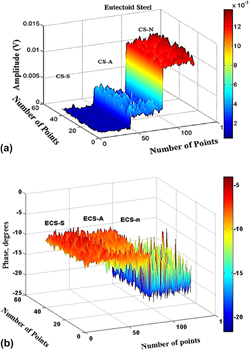
A eutectoid carbon steel was studied at three different annealing heat treatment cycles: spheroidizing, isothermal annealing, and normalization (air cooling). The aim of this study was to determine the correlation among thermal, structural, and metallurgical properties, as a result of the annealing heat treatment. Microstructure differences were produced by the heat treatment cooling rate with significant effects on Vickers nanohardness, thermal properties, and crystallinity. It was reflected in photothermal radiometry (PTR) images as in thermal conductivity and diffusivity. The amplitude signal increased as the cooling rate increased. It means that as the cooling rate increased, crystallinity, thermal diffusivity, and conductivity decreased. The cooling rate affected the metallurgical structure directly, and consequently, the nanohardness which decreased due to the solid solution formation and decomposition of the pearlite phase. As the cooling rate increased, the nanohardness increased modifying structural properties and the steel crystallinity. As the cooling rate decreased, the crystallinity increased.

Type
Articles
Copyright
Copyright © Materials Research Society 2017 

Access options

Get access to the full version of this content by using one of the access options below. (Log in options will check for institutional or personal access. Content may require purchase if you do not have access.)

Footnotes

Contributing Editor: Jürgen Eckert

References

REFERENCES

Zhou, D.S. and Shiflet, G.J.: Ferrite: Cementite crystallography in pearlite. Metall. Trans. A. 23(A), 1259 (1992).Google Scholar
Pitsch, W.: Der Orientierungszusammenhang zwischen Zementit und Ferrit im Perlit. Acta Metall. 10(1), 79 (1962).Google Scholar
Bowden, H.G. and Kelly, P.M.: Deformation twinning in shock-loaded pearlite. Acta Metall. 15(1), 105 (1967).Google Scholar
Boswell, P.G. and Chadwick, G.A.: The crystallography of herringbone pearlite. Scr. Metall. 11(11), 1001 (1977).CrossRefGoogle Scholar
Lara-Guevara, A., Ortiz-Echeverri, C.J., Rojas-Rodriguez, I., Mosquera-Mosquera, J.C., Ariza-Calderón, H., Ayala-Garcia, I., and Rodriguez-García, M.E.: Microstructural, structural, and thermal characterization of annealed carbon steels. Int. J. Thermophys. 37(99), 1 (2016).CrossRefGoogle Scholar
Busse, G.: Topics in Current Physics. In SPRINGER Series (Springer, Heidelberg, 1989); p. 251.Google Scholar
Busse, G. and Walther, H.G.: Progress of Photothermal and Photoacoustic Science and Technology, Vol. 1 (Elsevier, New York, 1992); p. 206.Google Scholar
Arai, T. and Harper, S.: ASM Handbook, Heat Treatment, Vol. 4 (ASM International, Novelty, 2006); p. 1012.Google Scholar
Bailey, A.R.: The Structure and Strength of Metals, Annotated Metallographic Specimens (Gatwick Press Ltd., Betchworth, 1967); pp. 4661.Google Scholar
Vander Voort, G.F.: Metallography and Microstructures ASM Handbook, Vol. 9 (ASM International, Novelty, 2004); pp. 103130.Google Scholar
Garcia, J.A., Guo, X., Mandelis, A., and Simmons, A.: Photo-Carrier-Radiometry (PCR) Metrology for Semiconductor Manufacturing Inspection, Vol. 788 (AIP Conf. Proc., Richardson, TX, USA, 2005); p. 625.Google Scholar
Garcia, J.A., Nicolaides, L., Park, P., Mandelis, A., and Farahkbahsh, B.: Photothermal radiometry of thermal sprayed coatings: Novel roughness elimination methodology. Anal. Sci. 17(1), 89 (2001).Google Scholar
Guo, X., Sivagurunathan, K., Garcia, J.A., Mandelis, A., Giunta, A., and Milletari, S.: Laser photothermal radiometric instrumentation for fast in-line industrial steel hardness inspection and case depth measurements. Appl. Opt. 48(C), 11 (2009).Google Scholar
Rojas-Rodriguez, I., Velazquez, R., Jaramillo, D., and Rodriguez-Garcia, M.E.: Correlation between chemical composition of silver alloys and photothermal radiometry signals. Int. J. Thermophis. 33(12), 2382 (2012).Google Scholar
Rosencwaig, A. and Gersho, A.: Theory of the photoacoustic effect with solids. J. Appl. Phys. 47(1), 64 (1976).CrossRefGoogle Scholar
Dias, D.T., Bedeschi, V.C., Ferreira da Silva, A., Nakamura, O., Castro Meira, M.V., and Trava-Airoldi, V.J.: Photoacoustic spectroscopy and thermal diffusivity measurement on hydrogenated amorphous carbon. Thin films deposited by plasma-enhanced chemical vapor deposition. Diamond Relat. Mater. 48(1), 1 (2014).Google Scholar
Yao, J. and Wang, L.V.: Sensitivity of photoacoustic microscopy. Photoacoustics 2(2), 87 (2014).Google Scholar
Marquenzini, M.V., Cell, N., Mansanares, A.M., Vargas, H., and Miranda, L.C.M.: Open photoacoustic cell spectroscopy. Meas. Sci. Technol. 2(1), 396 (1991).Google Scholar
Hawbolt, E.B., Brimacombe, J.K., and Chau, B.: Kinetics of austenite-pearlite transformation in eutectoid carbon steel. Metall. Mater. Trans. A 14(9), 1803 (1983).Google Scholar
Rojas-Rodriguez, I., Jaramillo-Vigueras, D., Velásquez-Hernández, R., del Real, A., Serroukh, I., Baños, L., García, J., and Rodriguez-García, M.E.: Thermal and structural characterization of copper-steel bonding interfaces produced by impact welding. Mater. Manuf. Processes 23(8), 823 (2008).Google Scholar
Ganesh, P., Nagpure, D.C., Gupta, R.K., and Kukreja, L.M.: Non-destructive micro-structural characterization of metallic specimens with a portable X-ray diffraction based residual stress analyzer. Stud. Eng. Technol. 2(1), 2330 (2015).Google Scholar
Swanson, H.E., Gilfrich, T., and Ugrinie, M.: Standard X-ray diffraction powder patterns. US Natl. Bur. Stand. 539(IV), 3 (1955).Google Scholar
Vashista, M. and Paul, S.: Correlation between full width at half maximum (FWHM) of XRD peak with residual stress on ground surfaces. Philos. Mag. 92(33), 4194 (2012).CrossRefGoogle Scholar
Moore, D.M. and Reynolds, R.C. Jr: X-ray Diffraction and the Identification and Analysis of Clay Minerals (University Press, Oxford, 1997); p. 332.Google Scholar