Hostname: page-component-848d4c4894-sjtt6 Total loading time: 0 Render date: 2024-06-18T22:43:14.615Z Has data issue: false hasContentIssue false

Analysis of in vitro corrosion behavior and hemocompatibility of electrophoretically deposited bioglass–chitosan–iron oxide coating for biomedical applications

Published online by Cambridge University Press:  22 June 2020

Sandeep Singh*
Affiliation:
Department of Mechanical Engineering, Punjabi University Patiala, Punjab, India
Gurpreet Singh
Affiliation:
Department of Mechanical Engineering, Punjabi University Patiala, Punjab, India
Niraj Bala
Affiliation:
Department of Mechanical Engineering, BBSBEC Fatehgarh Sahib, Punjab, India
*
a)Address all correspondence to this author. e-mail: sandeep_me@pbi.ac.in
Get access

Abstract

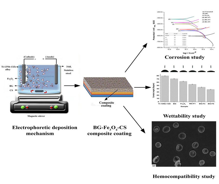
Electrophoretic deposition consisting of bioglass (BG)–chitosan (CS)–iron oxide nanoparticles (Fe3O4 NPs) on the Ti–13Nb–13Zr substrate was described. The bioactive coating was embedded in a CS matrix. The Fe3O4 NPs collected using the co-precipitation method varied at three different levels (1, 3, and 5 wt%) in the BG coating. The formulated coatings exhibited a hydrophilic character due to higher surface roughness values. The pull-off tape test was performed to check the adhesion strength of coatings. The composite coatings displayed adhesion strength of 5B class. The corrosion behavior was evaluated in Ringer's solution by the electrochemical test. The corrosion results showed that the composite coatings were more impressive as compared to pure BG and Fe3O4 coatings. The hemocompatibility results showed a hemolytic ratio (<5%), which validates them as favorable blood compatible nature of the deposited coatings. The findings exhibited that the BG–Fe3O4–CS coating can be widely employed as a favorable material for orthopedic applications.

Type
Article
Copyright
Copyright © Materials Research Society 2020

Access options

Get access to the full version of this content by using one of the access options below. (Log in options will check for institutional or personal access. Content may require purchase if you do not have access.)

References

Ananth, K.P., Suganya, K.S., Mangalaraj, D., Ferreira, J.M.F., and Balamurugan, A.: Electrophoretic bilayer deposition of zirconia and reinforced bioglass system on Ti-6Al-4V for implant applications: An in vitro investigation. Mater. Sci. Eng., C 33, 41604166 (2013).CrossRefGoogle Scholar
Balla, V.K., Bhat, A., Bose, S., and Bandyopadhyay, A.: Laser processed TiN reinforced Ti6Al4 V composite coatings. J. Mech. Behav. Biomed. 6, 920 (2012).CrossRefGoogle Scholar
Kaur, M. and Singh, K.: Review on titanium and titanium based alloys as biomaterials for orthopaedic applications. Mater. Sci. Eng., C 102, 844862 (2019).CrossRefGoogle ScholarPubMed
Jugowiec, D., Kot, M., and Moskalewicz, T.: Electrophoretic deposition and characterization of chitosan coatings on near-β titanium alloy. Arch. Metall. Mater. 61, 657664 (2016).CrossRefGoogle Scholar
He, Y., Zhang, Y., Jiang, Y., and Zhou, R.: Fabrication and characterization of superelastic Ti–Nb alloy enhanced with antimicrobial Cu via spark plasma sintering for biomedical applications. J. Mater. Res. 32, 25102520 (2017).CrossRefGoogle Scholar
Hu, H.Y., Zhang, L., He, Z.Y., Jiang, Y.H., and Tan, J.: Microstructure evolution, mechanical properties, and enhanced bioactivity of Ti-13Nb-13Zr based calcium pyrophosphate composites for biomedical applications. Mater. Sci. Eng., C 98, 279287 (2019).CrossRefGoogle ScholarPubMed
Dercz, G., Matuła, I., Zubko, M., and Dercz, J.: Phase composition and microstructure of new Ti–Ta–Nb–Zr biomedical alloys prepared by mechanical alloying method. Powder Diffr. 32, S186S192 (2017).CrossRefGoogle Scholar
Das, K., Bose, S., and Bandyopadhyay, A.: TiO2 nanotubes on Ti: Influence of nanoscale morphology on bone cell–materials interaction. J. Biomed. Mater. Res., A 90, 225237 (2009).CrossRefGoogle ScholarPubMed
Ke, Z., Yi, C., Zhang, L., He, Z.Y., Tan, J., and Jiang, Y.H.: Characterization of a new Ti-13Nb-13Zr-10Cu alloy with enhanced antibacterial activity for biomedicalapplications. Mater. Lett. 253, 335338 (2019).CrossRefGoogle Scholar
Roy, M., Balla, V.K., Bandyopadhyay, A., and Bose, S.: MgO-doped tantalum coating on Ti: Microstructural study and biocompatibility evaluation. ACS Appl. Mater. Interfaces 4, 577580 (2012).CrossRefGoogle ScholarPubMed
López-Cuevas, J., Rendón-Angeles, J.C., Méndez-Nonell, J., and Barrientos-Rodríguez, H.: In vitro bioactivity of AISI 316L stainless steel coated with hydroxyapatite-seeded 58S bioglass. MRS Adv. 4, 31333142 (2019).CrossRefGoogle Scholar
Mahlooji, E., Atapour, M., and Labbaf, S.: Electrophoretic deposition of bioactive glass–Chitosan nanocomposite coatings on Ti-6Al-4V for orthopedic applications. Carbohydr. Polym. 226, 112 (2019).CrossRefGoogle Scholar
Hench, L.L.: Biomaterials: A forecast for the future. Biomaterials 19, 14191423 (1998).CrossRefGoogle ScholarPubMed
Esteban, S.L., Saiz, E., Fujino, S., Oku, T., Suganuma, K., and Tomsia, A.P.: Bioactive glass coatings for orthopedic metallic implants. J. Eur. Ceram. Soc. 23, 29212930 (2003).CrossRefGoogle Scholar
Fu, Q., Saiz, E., Rahaman, M.N., and Tomsia, A.P.: Bioactive glass scaffolds for bone tissue engineering: state of the art and future perspectives. Mater. Sci. Eng., C 31, 12451256 (2011).CrossRefGoogle ScholarPubMed
Krause, D., Thomas, B., Leinenbach, C., Eifler, D., Minay, E.J., and Boccaccini, A.R.: The electrophoretic deposition of Bioglass particles on stainless steel and nitinol substrates. Surf. Coat. Technol. 200, 48354845 (2006).CrossRefGoogle Scholar
Castelló, M.E., Anbinder, P.S., Amalvy, J.I., and Peruzzo, P.J.: Production and characterization of chitosan and glycerol-chitosan films. MRS Adv. 3, 36013610 (2018).CrossRefGoogle Scholar
Jiang, T., Zhang, Z., Zhou, Y., Liu, Y., Wang, Z., Tong, H., Shen, X., and Wang, Y.: Surface functionalization of titanium with chitosan/gelatin via electrophoretic deposition: Characterization and cell behavior. Biomacromolecules 11, 12541260 (2010).CrossRefGoogle ScholarPubMed
Dong, P., Hao, W., Xia, Y., Da, G., and Wang, T.: Comparison study of corrosion behavior and biocompatibility of polyethyleneimine (PEI)/heparin and chitosan/heparin coatings on NiTi alloy. J. Mater. Sci. Technol. 26, 10271031 (2010).CrossRefGoogle Scholar
Martin, H.J., Schulz, K.H., Bumgardner, J.D., and Judith, A.: Schneider: Enhanced bonding of chitosan to implant quality titanium via four treatment combinations. Thin Solid Films 516, 62776286 (2008).CrossRefGoogle Scholar
Bumgardner, J.D., Wiser, R., Gerard, P.D., Bergin, P., Chestnutt, B., Marini, M., Ramsey, V., Elder, S.H., and Gilbert, J.A.: Chitosan: Potential use as a bioactive coating for orthopaedic and craniofacial/dental implants. J. Biomater. Sci., Polym. Ed. 14, 423438 (2003).CrossRefGoogle ScholarPubMed
Zhang, C., He, Y., Xu, Z., Shi, H., Di, H., Pan, Y., and Xu, W.: Fabrication of Fe3O4@SiO2 nanocomposites to enhance anticorrosion performance of epoxy coatings. Polym. Adv. Technol. 27, 740747 (2016).CrossRefGoogle Scholar
Mahdavi, M., Ahmad, M.B., Haron, M.J., Namvar, F., Nadi, B., Ab Rahman, M.Z., and Amin, J.: Synthesis, surface modification and characterisation of biocompatible magnetic iron oxide nanoparticles for biomedical applications. Molecules 18, 75337548 (2013).CrossRefGoogle ScholarPubMed
Huang, X., Hou, X., Zhang, X., Rosso, K.M., and Zhang, L.: Facet-dependent contaminant removal properties of hematite nanocrystals and their environmental implications. Environ. Sci.: Nano 5, 17901806 (2018).Google Scholar
Tran, N. and Webster, T.J.: Magnetic nanoparticles: Biomedical applications and challenges. J. Mater. Chem. 20, 87608767 (2010).CrossRefGoogle Scholar
Charoensuk, T., Sirisathitkul, C., Boonyang, U., Macha, I.J., Santos, J., Grossin, D., and Nissan, B.B.: In vitro bioactivity and stem cells attachment of three-dimensionally ordered macroporous bioactive glass incorporating iron oxides. J. Non-Cryst. Solids 452, 6273 (2016).CrossRefGoogle Scholar
Meliegy, E.E., Mabrouk, M., El-Sayed, S.A.M., Abd El-Hady, B.M., Shehata, M.R., and Hosny, W.M.: Novel Fe2O3-doped glass/chitosan scaffolds for bone tissue replacement. Ceram. Int. 44, 91409151 (2018).CrossRefGoogle Scholar
Wu, C., Fan, W., Zhu, Y., Gelinsky, M., Chang, J., Cuniberti, G., Albrecht, V., Friis, T., and Xiao, Y.: Multifunctional magnetic mesoporous bioactive glass scaffolds with a hierarchical pore structure. Acta Biomater. 7, 35633572 (2011).CrossRefGoogle ScholarPubMed
Martín-Saavedra, F.M., Hernández, E.R., Boré, A., Arcos, D., Regí, M.V., and Vilaboa, N.: Magnetic mesoporous silica spheres for hyperthermia therapy. Acta Biomater. 6, 45224531 (2010).CrossRefGoogle ScholarPubMed
Zhuang, J., Lin, S., Dong, L., Cheng, K., and Weng, W.: Magnetically actuated mechanical stimuli on Fe3O4/mineralized collagen coatings to enhance osteogenic differentiation of the MC3T3-E1 cells. Acta Biomater. 71, 4960 (2018).CrossRefGoogle ScholarPubMed
Sun, J., Zhou, S., Hou, P., Yang, Y., Weng, J., Li, X., and Li, M.: Synthesis and characterization of biocompatible Fe3O4 nanoparticles. J. Biomed. Mater. Res., A 80, 333341 (2007).CrossRefGoogle ScholarPubMed
Moskalewicz, T., Łukaszczyk, A., Kruk, A., Kot, M., Jugowiec, D., Dubiel, B., and Radziszewska, A.: Porous HA and nanocomposite nc-TiO2/HA coatings to improve the electrochemical corrosion resistance of the Co-28Cr-5Mo alloy. Mater. Chem. Phys. 199, 144158 (2017).CrossRefGoogle Scholar
Wu, C., Ramaswamy, Y., Gale, D., Yang, W., Xiao, K., Zhang, L., Yin, Y., and Zreiqat, H.: Novel sphene coatings on Ti–6Al–4 V for orthopedic implants using sol-gel method. Acta Biomater. 4, 569576 (2008).CrossRefGoogle Scholar
Fielding, G.A., Roy, M., Bandyopadhyay, A., and Bose, S.: Antibacterial and biological characteristics of silver containing and strontium doped plasma sprayed hydroxyapatite coatings. Acta Biomater. 8, 31443152 (2012).CrossRefGoogle ScholarPubMed
Roy, M., Bandyopadhyay, A., and Bose, S.: Laser surface modification of electrophoretically deposited hydroxyapatite coating on titanium. J. Am. Ceram. Soc. 91, 35173521 (2008).CrossRefGoogle Scholar
Yao, Z., Gao, H., Jiang, Z., and Wang, F.: Structure and properties of ZrO2 ceramic coatings on AZ91D Mg alloy by plasma electrolytic oxidation. J. Am. Ceram. Soc. 91, 555558 (2008).CrossRefGoogle Scholar
Uchikoshi, T., Ozawa, K., Hatton, B.D., and Sakka, Y.: Dense, bubble-free ceramic deposits from aqueous suspensions by electrophoretic deposition. J. Mater. Res. 16, 321324 (2001).CrossRefGoogle Scholar
Wang, C.A., Long, B., Lin, W., Huang, Y., and Sun, J.: Poly (amic acid)–clay nacrelike composites prepared by electrophoretic deposition. J. Mater. Res. 23, 17061712 (2008).CrossRefGoogle Scholar
Khanali, O., Baghshahi, S., and Rajabi, M.: Fabrication and characterization of YSZ/Al2O3 nano-composite coatings on Inconel by electrophoretic deposition. J. Mater. Res. 32, 34023408 (2017).CrossRefGoogle Scholar
Aghajani, H. and Pouzesh, M.: Electrophoretic deposition and corrosion behavior study of aluminum coating on AZ91D substrate. J. Particle Sci. Technol. 3, 219232 (2017).Google Scholar
Zhang, X., Li, X.W., Li, J.G., and Sun, X.D.: Preparation and characterizations of bioglass ceramic cement/Ca–P coating on pure magnesium for biomedical applications. ACS Appl. Mater. Interfaces 6, 513525 (2013).CrossRefGoogle ScholarPubMed
Sarkar, P. and Nicholson, P.S.: Electrophoretic deposition (EPD): Mechanisms, kinetics, and application to ceramics. J. Am. Ceram. Soc. 79, 19872002 (1996).CrossRefGoogle Scholar
Rojaee, R., Fathi, M., and Raeissir, K.: Cormparing nanostructured hydroxyapatite coating on AZ91 alloy samples via sol-gel and electrophoretic deposition for biomedical applications. IEEE Trans. Nanobiosci. 13, 409414 (2014).CrossRefGoogle ScholarPubMed
Heise, S., Höhlinger, M., Hernández, Y.T., Palacio, J.J.P., Ortiz, J.A.R., Wagener, V., Virtanen, S., and Boccaccini, A.R.: Electrophoretic deposition and characterization of chitosan/bioactive glass composite coatings on Mg alloy substrates. Electrochim. Acta 232, 456464 (2017).CrossRefGoogle Scholar
Mohammadi, R., Ordikhani, F., and Fray, D.J.: Template-based growth of titanium dioxide nanorods by a particulate sol-electrophoretic deposition process. Particuology 9, 161169 (2011).CrossRefGoogle Scholar
Agarwal, S., Curtin, J., Duffy, B., and Jaiswal, S.: Biodegradable magnesium alloys for orthopaedic applications: A review on corrosion, biocompatibility and surface modifications. Mater. Sci. Eng., C 68, 948963 (2016).CrossRefGoogle ScholarPubMed
Huang, K., Cai, S., Xu, G., Ye, X., Dou, Y., Ren, M., and Wang, X.: Preparation and characterization of mesoporous 45S5 bioactive glass–ceramic coatings on magnesium alloy for corrosion protection. J. Alloys Compd. 580, 290297 (2013).CrossRefGoogle Scholar
Salimkhani, H., Alanagh, F.M., Aghajani, H., and Bostanabad, K.O.: Study on the magnetic and microwave properties of electrophoretically deposited nano-Fe3O4 on carbon fiber. Proc. Mater. Sci. 11, 231237 (2015).CrossRefGoogle Scholar
Bajpai, I., Balani, K., and Basu, B.: Spark plasma sintered HA-Fe3O4 based multifunctional magnetic biocomposites. J. Am. Ceram. Soc. 96, 21002108 (2013).CrossRefGoogle Scholar
Molaei, A., Yari, M., and Reza Afshar, M.: Modification of electrophoretic deposition of chitosan–bioactive glass–hydroxyapatite nanocomposite coatings for orthopedic applications by changing voltage and deposition time. Ceram. Int. 41, 1453714544 (2015).CrossRefGoogle Scholar
Hou, P., Shi, C., Wu, L., and Hou, X.: Chitosan/hydroxyapatite/Fe3O4 magnetic composite for metal-complex dye AY220 removal: Recyclable metal-promoted Fenton-like degradation. Microchem. J. 128, 218225 (2016).CrossRefGoogle Scholar
Pishbin, F., Mouriño, V., Flor, S., Kreppel, S., Salih, V., Ryan, M.P., and Boccaccini, A.R.: Electrophoretic deposition of gentamicin-loaded bioactive glass/chitosan composite coatings for orthopaedic implants. ACS Appl. Mater. Interfaces 6, 87968806 (2014).CrossRefGoogle ScholarPubMed
Caridad, G.S., Merino, E.G., Alves, N.M., Bermudez, V.D.Z., Boccaccini, A.R., and Mano, J.F.: Chitosan membranes containing micro or nano-size bioactive glass particles: Evolution of biomineralization followed by in situ dynamic mechanical analysis. J. Mech. Behav. Biomed. Mater. 20, 173183 (2013).CrossRefGoogle Scholar
Mehdipour, M. and Afshar, A.: A study of the electrophoretic deposition of bioactive glass–chitosan composite coating. Ceram. Int. 38, 471476 (2012).CrossRefGoogle Scholar
Guo, Y., Zhou, Y., Jia, D., and Meng, Q.: Fabrication and in vitro characterization of magnetic hydroxycarbonate apatite coatings with hierarchically porous structures. Acta Biomater. 4, 923931 (2008).CrossRefGoogle ScholarPubMed
Mohan, L., Durgalakshmi, D., Geetha, M., Sankara Narayanan, T.S.N., and Asokamani, R.: Electrophoretic deposition of nanocomposite (HAp + TiO2) on titanium alloy for biomedical applications. Ceram. Int. 38, 34353443 (2012).CrossRefGoogle Scholar
Rivera, L.R., Distaso, M., Peukert, W., and Boccaccini, A.R.: Electrophoretic deposition of anisotropic α-Fe2O3/PVP/chitosan nanocomposites for biomedical applications. Mater. Lett. 200, 8386 (2017).CrossRefGoogle Scholar
Yona, K.S., Rittel, D., and Dorogoy, A.: Mechanical assessment of grit blasting surface treatments of dental implants. J. Mech. Behav. Biomed. Mater. 39, 375390 (2014).CrossRefGoogle Scholar
Hong, W., Guo, F., Chen, J., Wang, X., Zhao, X., and Xiao, P.: Bioactive glass–chitosan composite coatings on PEEK: Effects of surface wettability and roughness on the interfacial fracture resistance and in vitro cell response. Appl. Surf. Sci. 440, 514523 (2018).CrossRefGoogle Scholar
Deligianni, D.D., Katsala, N.D., Koutsoukos, P.G., and Missirlis, Y.F.: Effect of surface roughness of hydroxyapatite on human bone marrow cell adhesion, proliferation, differentiation and detachment strength. Biomaterials 22, 8796 (2000).CrossRefGoogle Scholar
Padial-Molina, M., Galindo-Moreno, P., Fernández-Barbero, J.E., O'Valle, F., Jódar-Reyes, A.B., Ortega-Vinuesa, J.L., and Ramón-Torregrosa, P.J.: Role of wettability and nanoroughness on interactions between osteoblast and modified silicon surfaces. Acta Biomater. 7, 771778 (2011).CrossRefGoogle ScholarPubMed
Gehroudi, B., Could, T.R.L., and Brunette, D.M.: Titanium coated micromachined grooves of different dimensions effect epithelial and connective tissue cells differently in vivo. J. Biomed. Mater. Res. 24, 12031219 (1990).Google Scholar
Singh, S., Singh, G., and Bala, N.: Electrophoretic deposition of hydroxyapatite-iron oxide-chitosan composite coatings on Ti-13Nb-13Zr alloy for biomedical applications. Thin Solid Films 697, 137801137812 (2020).CrossRefGoogle Scholar
Menzies, K.L. and Jones, L.: The impact of contact angle on the biocompatibility of biomaterials. Optom. Vis. Sci. 87, 387399 (2010).Google ScholarPubMed
Chau, T.T., Bruckard, W.J., Koh, P.T.L., and Nguyen, A.V.: A review of factors that affect contact angle and implications for flotation practice. Adv. Colloid Interface Sci. 150, 106115 (2009).CrossRefGoogle ScholarPubMed
Kylián, O., Polonskyi, O., Kratochvíl, J., Artemenko, A., Choukourov, A., Drábik, M., Solař, P., Slavínská, D., and Biederman, H.: Control of wettability of plasma polymers by application of Ti nano-clusters. Plasma Process. Polym. 9, 180187 (2012).CrossRefGoogle Scholar
Rattan, P.C., Singh, B.P., Besra, L., and Bhattacharjee, S.: Multiwalled carbon nanotubes reinforced hydroxyapatite-chitosan composite coating on Ti metal: Corrosion and mechanical properties. J. Am. Ceram. Soc. 95, 27252731 (2012).Google Scholar
Göncü, Y., Geçgin, M., Bakan, F., and Ay, N.: Electrophoretic deposition of hydroxyapatite-hexagonal boron nitride composite coatings on Ti substrate. Mater. Sci. Eng., C 79, 343353 (2017).CrossRefGoogle ScholarPubMed
Molaei, A., Amadeh, A., Yari, M., and Afshar, M.R.: Structure, apatite-inducing ability and corrosion behavior of chitosan/halloysite nanotube coatings prepared by electrophoretic deposition on titanium substrate. Mater. Sci. Eng., C 59, 740747 (2016).CrossRefGoogle ScholarPubMed
Sharma, S., Soni, V.P., and Bellare, J.R.: Chitosan reinforced apatite–wollastonite coating by electrophoretic deposition on titanium implants. J. Mater. Sci. Mater. Med. 20, 14271436 (2009).CrossRefGoogle ScholarPubMed
Unsoy, G., Yalcin, S., Khodadust, R., Gunduz, G., and Gunduz, U.: Synthesis optimization and characterization of chitosan-coated iron oxide nanoparticles produced for biomedical applications. J. Nanopart. Res. 14, 964–698 (2012).CrossRefGoogle Scholar
Manam, N.S., Harun, W.S.W., Shri, D.N.A., Ghani, D.S.A.C., Kurniawa, T., Ismail, M.H., and Ibrahim, M.H.I.: Study of corrosion in biocompatible metals for implants: A review. J. Alloys Compd. 701, 698715 (2017).CrossRefGoogle Scholar
Shikha, D., Shahid, M., Jha, U., Sinha, S.K., Reddy, V.R., Ojha, S., Kumar, P., and Kanjilal, D.: Corrosion, wettability and thrombogenicity investigation of ion beam modified HA/Al2O3. Mater. Chem. Phys. 163, 272278 (2015).CrossRefGoogle Scholar
Poorraeisi, M. and Afshar, A.: The study of electrodeposition of hydroxyapatite-ZrO2-TiO2 nanocomposite coatings on 316 stainless steel. Surf. Coat. Technol. 339, 199207 (2018).CrossRefGoogle Scholar
Jugowiec, D., Lukaszczyk, A., Cienick, L., and Mokalewicz, T.: Electrophoretic deposition and characterization of composite chitosan based coatings incorporating bioglass and sol-gel glass particle on the Ti-13Nb-13Zr alloy. Surf. Coat. Technol. 17, 3346 (2017).CrossRefGoogle Scholar
Kiahosseini, S.R., Afshar, A., Larijani, M.M., and Yousefpour, M.: Structural and corrosion characterization of hydroxyapatite/zirconium nitride-coated AZ91 magnesium alloy by ion beam sputtering. App. Surf. Sci. 401, 172180 (2017).CrossRefGoogle Scholar
Arias, L.C., Polo, S.C., Gao, H., Gilabert, J., Sanchez, E., Roether, J.A., Schubert, D.W., Virtanen, S., and Boccaccini, A.R.: Electrophoretic deposition of nanostructured-TiO2/chitosan composite coatings on stainless steel. RSC Adv. 28, 1124711254 (2015).Google Scholar
Zhang, S., Li, J., Song, Y., Zhao, C., Zhang, X., Xie, C., Zhang, Y., Tao, H., He, Y., Jiang, Y., and Bian, Y.: In vitro degradation, hemolysis and MC3T3-E1 cell adhesion of biodegradable Mg–Zn alloy. Mater. Sci. Eng., C 29, 19071912 (2009).CrossRefGoogle Scholar
Henkelman, S., Rakhorst, G., Blanton, J., and Oeveren, W.V.: Standardization of incubation conditions for hemolysis testing of biomaterials. Mater. Sci. Eng., C 29, 16501654 (2009).CrossRefGoogle Scholar
Anjaneyulu, U., Swaroop, V.K., and Vijayalakshmi, U.: Preparation and characterization of novel Ag-doped hydroxyapatite–Fe3O4–chitosan hybrid composites and in vitro biological evaluations for orthopedic applications. RSC Adv. 13, 1099711007 (2016).CrossRefGoogle Scholar
Heidari, F., Razavi, M., Bahrololoom, M.E., Yazdimamaghani, M., Tahriri, M., Kotturi, H., and Tayebi, L.: Evaluation of the mechanical properties, in vitro biodegradability and cytocompatibility of natural chitosan/hydroxyapatite/nano-Fe3O4 composite. Ceram. Int. 44, 275281 (2018).CrossRefGoogle Scholar
Molaei, A., Yari, M., and Afshar, M.R.: Investigation of halloysite nanotube content on electrophoretic deposition (EPD) of chitosan-bioglass-hydroxyapatite-halloysite nanotube nanocomposites films in surface engineering. Appl. Clay Sci. 135, 7581 (2017).CrossRefGoogle Scholar
Besra, L. and Liu, M.: A review on fundamentals and applications of electrophoretic deposition (EPD). Prog. Mater. Sci. 52, 161 (2007).CrossRefGoogle Scholar
Wang, Z., Shemilt, J., and Xiao, P.: Fabrication of ceramic composite coatings using electrophoretic deposition, reaction bonding and low temperature sintering. J. Eur. Ceram. Soc. 22, 183189 (2002).CrossRefGoogle Scholar
Li, B., Niu, J., Liu, H., and Li, G.: Fabrication and corrosion property of novel 3-aminopropyltriethoxy-modified calcium phosphate/poly (lactic acid) composite coating on AZ60 Mg alloy. Appl. Phys. A: Mater. Sci. Process. 124, 113 (2018).CrossRefGoogle Scholar
Lakshmi, R.V. and Basu, B.J.: Fabrication of superhydrophobic sol–gel composite films using hydrophobically modified colloidal zinc hydroxide. J. Colloid Interface Sci. 339, 454460 (2009).CrossRefGoogle ScholarPubMed
Dora, C.P., Kushwah, V., Katiyar, S.S., Kumar, P., Pillay, V., Suresh, S., and Jain, S.: Improved metabolic stability and therapeutic efficacy of a novel molecular gemcitabine phospholipid complex. Int. J. Pharm. 530, 113127 (2017).CrossRefGoogle ScholarPubMed
Singh, B., Singh, G., Sidhu, B.S., and Bhatia, N.: In-vitro assessment of HA-Nb coating on Mg alloy ZK60 for biomedical applications. Mater. Chem. Phys. 231, 138149 (2019).CrossRefGoogle Scholar