Hostname: page-component-78c5997874-j824f Total loading time: 0 Render date: 2024-11-17T21:48:44.077Z Has data issue: false hasContentIssue false

Highly fluorescent CdTe nanocrystals: Synthesis, characterization, property, mechanism, and application as a sensor for biomolecule analysis

Published online by Cambridge University Press:  04 March 2014

Jun Yao
Affiliation:
Research Center of Analytical Instrumentation, Analytical and Testing Center, College of Chemistry, Sichuan University, Chengdu, Sichuan 610064, People's Republic of China
Mei Yang
Affiliation:
Research Center of Analytical Instrumentation, Analytical and Testing Center, College of Chemistry, Sichuan University, Chengdu, Sichuan 610064, People's Republic of China
Yixiang Duan*
Affiliation:
Research Center of Analytical Instrumentation, Analytical and Testing Center, College of Chemistry, Sichuan University, Chengdu, Sichuan 610064, People's Republic of China
*
a)Address all correspondence to this author. e-mail: yduan@scu.edu.cn
Get access

Abstract

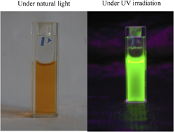
Highly luminescent CdTe quantum dots (QDs) were prepared through a fast, facile, and straightforward method. The crystal structure, particle size, optical properties as well as molecular interactions between the CdTe QDs and their capping agents have been investigated by high resolution transmission electron microscopy, selected area electron diffraction, scanning transmission electron microscope–energy dispersive x-ray spectroscopy, UV-vis absorption, photoluminescence, and Fourier transform infrared, respectively. The results illustrate that the CdTe nanoparticles exhibit cubic structure and the average crystallite size is 2.3 nm. Meanwhile, fluorescence and UV-vis spectroscopic techniques were used to study the interaction between hemin and the well-defined CdTe QDs. In weak basic media, the fluorescence of CdTe QDs was quenched notably by hemin, and the quenching values were proportional to the concentration of the quencher in a certain range. The quenching mechanism was discussed to be a dynamic quenching procedure, collisional process, and hemin as a fluorescence quencher donated its electron to CdTe QDs to occupy the hole and accordingly disrupted the electron–hole recombination.

Type
Articles
Copyright
Copyright © Materials Research Society 2014 

Access options

Get access to the full version of this content by using one of the access options below. (Log in options will check for institutional or personal access. Content may require purchase if you do not have access.)

References

REFERENCES

Yao, J., Sun, Y., Yang, M., and Duan, Y.: Chemistry, physics and biology of graphene-based nanomaterials: New horizons for sensing, imaging and medicine. J. Mater. Chem. 22(29), 14313 (2012).CrossRefGoogle Scholar
Yang, M., Yao, J., and Duan, Y.: Graphene and its derivatives for cell biotechnology. Analyst 138(1), 72 (2013).CrossRefGoogle ScholarPubMed
Kamat, P.V., Tvrdy, K., Baker, D.R., and Radich, J.G.: Beyond photovoltaics: Semiconductor nanoarchitectures for liquid-junction solar cells. Chem. Rev. 110(11), 6664 (2010).CrossRefGoogle ScholarPubMed
Medintz, I.L., Stewart, M.H., Trammell, S.A., Susumu, K., Delehanty, J.B., Mei, B.C., Melinger, J.S., Blanco-Canosa, J.B., Dawson, P.E., and Mattoussi, H.: Quantum-dot/dopamine bioconjugates function as redox coupled assemblies for in vitro and intracellular pH sensing. Nat. Mater. 9(8), 676 (2010).CrossRefGoogle ScholarPubMed
Wu, L., Quan, B., Liu, Y., Song, R., and Tang, Z.: One-pot synthesis of liquid Hg/solid beta-HgS metal-semiconductor heterostructures with unique electrical properties. ACS Nano 5(3), 2224 (2011).CrossRefGoogle ScholarPubMed
Chaudhuri, R.G. and Paria, S.: Core/shell nanoparticles: Classes, properties, synthesis mechanisms, characterization, and applications. Chem. Rev. 112(4), 2373 (2012).CrossRefGoogle Scholar
So, M.K., Xu, C.J., Loening, A.M., Gambhir, S.S., and Rao, J.H.: Self-illuminating quantum dot conjugates for in vivo imaging. Nat. Biotechnol. 24(3), 339 (2006).CrossRefGoogle ScholarPubMed
Wu, X.Y., Liu, H.J., Liu, J.Q., Haley, K.N., Treadway, J.A., Larson, J.P., Ge, N., Peale, F., and Bruchez, M.P.: Immunofluorescent labeling of cancer marker Her2 and other cellular targets with semiconductor quantum dots. Nat. Biotechnol. 21(1), 41 (2003).CrossRefGoogle ScholarPubMed
Mattoussi, H., Mauro, J.M., Goldman, E.R., Anderson, G.P., Sundar, V.C., Mikulec, F.V., and Bawendi, M.G.: Self-assembly of CdSe-ZnS quantum dot bioconjugates using an engineered recombinant protein. J. Am. Chem. Soc. 122(49), 12142 (2000).CrossRefGoogle Scholar
Nozik, A.J., Beard, M.C., Luther, J.M., Law, M., Ellingson, R.J., and Johnson, J.C.: Semiconductor quantum dots and quantum dot arrays and applications of multiple exciton generation to third-generation photovoltaic solar cells. Chem. Rev. 110(11), 6873 (2010).CrossRefGoogle Scholar
Samia, A.C.S., Chen, X.B., and Burda, C.: Semiconductor quantum dots for photodynamic therapy. J. Am. Chem. Soc. 125(51), 15736 (2003).CrossRefGoogle ScholarPubMed
Shi, Y., Wang, J., Li, S., Wang, Z., Zang, X., Zu, X., Zhang, X., Guo, F., and Tong, G.: Photoluminescence-enhanced CdTe quantum dots by hyperbranched poly (amidoamine) s functionalization. J. Mater. Res. 28(14), 1940 (2013).CrossRefGoogle Scholar
Gao, X.H. and Nie, S.M.: Doping mesoporous materials with multicolor quantum dots. J. Phys. Chem. B 107(42), 11575 (2003).CrossRefGoogle Scholar
Gao, X.H. and Nie, S.M.: Quantum dot-encoded mesoporous beads with high brightness and uniformity: Rapid readout using flow cytometry. Anal. Chem. 76(8), 2406 (2004).CrossRefGoogle ScholarPubMed
Aragay, G., Pino, F., and Merkoçi, A.: Nanomaterials for sensing and destroying pesticides. Chem. Rev. 112(10), 5317 (2012).CrossRefGoogle ScholarPubMed
Zhou, H., Zhou, G., Du, Q., Bi, H., and Zhou, J.: Surfactant-assisted reflux synthesis of PbS nanostructures and their properties. J. Mater. Res. 1(1), 1 (2012).Google Scholar
Jaiswal, J.K., Mattoussi, H., Mauro, J.M., and Simon, S.M.: Long-term multiple color imaging of live cells using quantum dot bioconjugates. Nat. Biotechnol. 21(1), 47 (2003).CrossRefGoogle ScholarPubMed
Somers, R.C., Bawendi, M.G., and Nocera, D.G.: CdSe nanocrystal based chem-/bio-sensors. Chem. Soc. Rev. 36(4), 579 (2007).CrossRefGoogle ScholarPubMed
Dubertret, B., Skourides, P., Norris, D.J., Noireaux, V., Brivanlou, A.H., and Libchaber, A.: In vivo imaging of quantum dots encapsulated in phospholipid micelles. Science 298(5599), 1759 (2002).CrossRefGoogle ScholarPubMed
Kobayashi, H., Ogawa, M., Alford, R., Choyke, P.L., and Urano, Y.: New strategies for fluorescent probe design in medical diagnostic imaging. Chem. Rev. 110(5), 2620 (2010).CrossRefGoogle ScholarPubMed
Chen, X-F., Zhou, M., Chang, Y-P., Ren, C-L., Chen, H-L., and Chen, X-G.: Novel synthesis of beta-cyclodextrin functionalized CdTe quantum dots as luminescent probes. Appl. Surf. Sci. 263, 491 (2012).CrossRefGoogle Scholar
Qiao, X., Jian-Hao, W., Zhan, W., Zhao-Hui, Y., Qin, Y., and Yuan-Di, Z.: Interaction of CdTe quantum dots with DNA. Electrochem. Commun. 10(9), 1337 (2008).Google Scholar
Callan, J.F., Mulrooney, R.C., Kamila, S., and McCaughan, B.: Anion sensing with luminescent quantum dots: A modular approach based on the photoinduced electron transfer (PET) mechanism. J. Fluoresc. 18(2), 527 (2008).CrossRefGoogle ScholarPubMed
Neuman, D., Ostrowski, A.D., Mikhailovsky, A.A., Absalonson, R.O., Strouse, G.F., and Ford, P.C.: Quantum dot fluorescence quenching pathways with Cr(III) complexes. Photosensitized NO production from trans-Cr(cyclam)(ONO)(2)(+) J. Am. Chem. Soc. 130(1), 168 (2008).CrossRefGoogle Scholar
Jin, T., Fujii, F., Yamada, E., Nodasaka, Y., and Kinjo, M.: Preparation and characterization of thiacalix[4]arene coated water-soluble CdSe/ZnS quantum dots as a fluorescent probe for Cu2+ ions. Comb. Chem. High Throughput Screening 10(6), 473 (2007).CrossRefGoogle ScholarPubMed
Wang, Q., Yang, L., Fang, T., Wu, S., Liu, P., Min, X., and Li, X.: Interactions between CdSe/CdS quantum dots and DNA through spectroscopic and electrochemical methods. Appl. Surf. Sci. 257(23), 9747 (2011).CrossRefGoogle Scholar
Tsiftsoglou, A.S., Tsamadou, A.I., and Papadopoulou, L.C.: Heme as key regulator of major mammalian cellular functions: Molecular, cellular, and pharmacological aspects. Pharmacol. Ther. 111(2), 327 (2006).CrossRefGoogle ScholarPubMed
Lu, N., Yi, L., Deng, Q., Li, J., Gao, Z., and Li, H.: The interaction between desferrioxamine and hemin: A potential toxicological implication. Toxicol. In Vitro 26(5), 732 (2012).CrossRefGoogle ScholarPubMed
Lu, N., Zhang, M., Li, H., and Gao, Z.: Completely different effects of desferrioxamine on hemin/nitrite/H2O2-induced bovine serum albumin nitration and oxidation. Chem. Res. Toxicol. 21(6), 1229 (2008).CrossRefGoogle ScholarPubMed
Yu, W.W., Qu, L., Guo, W., and Peng, X.: Experimental determination of the extinction coefficient of CdTe, CdSe, and CdS nanocrystals. Chem. Mater. 15(14), 2854 (2003).CrossRefGoogle Scholar
Colthup, N., Daly, L., and Wiberley, S.: Introduction to Infrared and Raman Spectroscopy (Hacourt Brace Jovanovich, 1990), p. 291.Google Scholar
Han, S.W., Han, H.S., and Kim, K.: Infrared and Raman spectra of 4-cyanobenzoic acid on powdered silver. Vib. Spectrosc. 21(1–2), 133 (1999).CrossRefGoogle Scholar
Tao, Y.T.: Structural comparison of self-assembled monolayers of n-alkanoic acids on the surfaces of silver, copper, and aluminum. J. Am. Chem. Soc. 115(10), 4350 (1993).CrossRefGoogle Scholar
Wang, S-G., Yang, Q-B., Bai, J., Song, Y., Zhang, C-Q., and Li, Y-X.: Transferring CdTe nanoparticles from liquid phase to polyvinylpyrrolidone nanofibers by electrospinning and detecting its photoluminescence property. Chem. Res. Chin. Univ. 24(4), 459 (2008).CrossRefGoogle Scholar
Zhang, H., Zhou, Z., Yang, B., and Gao, M.: The influence of carboxyl groups on the photoluminescence of mercaptocarboxylic acid-stabilized CdTe nanoparticles. J. Phys. Chem. B 107(1), 8 (2003).CrossRefGoogle Scholar
Huang, D., Geng, F., Liu, Y., Wang, X., Jiao, J., and Yu, L.: Biomimetic interactions of proteins with functionalized cadmium sulfide quantum dots. Colloids Surf., A 392(1), 191 (2011).CrossRefGoogle Scholar
Xiao, Q., Huang, S., Qi, Z-D., Zhou, B., He, Z-K., and Liu, Y.: Conformation, thermodynamics and stoichiometry of HSA adsorbed to colloidal CdSe/ZnS quantum dots. Biochim. Biophys. Acta 1784(7), 1020 (2008).CrossRefGoogle ScholarPubMed
Peng, J., Liu, S., Yan, S., Fan, X., and He, Y.: A study on the interaction between CdTe quantum dots and chymotrypsin using optical spectroscopy. Colloids Surf., A 359, 13 (2010).CrossRefGoogle Scholar
Chen, L., Zhang, X. , Zhang, C., Zhou, G., Zhang, W., Xiang, D., He, Z., and Wang, H.: Dual-color fluorescence and homogeneous immunoassay for the determination of human enterovirus 71. Anal. Chem. 83, 7316 (2011).CrossRefGoogle ScholarPubMed
Comby, S. and Gunnlaugsson, T.: Luminescent lanthanide-functionalized gold nanoparticles: Exploiting the interaction with bovine serum albumin for potential sensing applications. ACS Nano. 5(9), 7184 (2011).CrossRefGoogle ScholarPubMed
Mikhail, B.Y. and Achilefu, S.: Fluorescence lifetime measurements and biological imaging. Chem. Rev. 110, 2641 (2010).Google Scholar
Lakowicz, J.R.: Principles of Fluorescence Spectroscopy (Springer, New York, NY, 2009).Google Scholar
Azzazy, H.M.E., Mansour, M.M.H., and Kazmierczak, S.C.: From diagnostics to therapy: Prospects of quantum dots. Clin. Biochem. 40(13–14), 917 (2007).CrossRefGoogle ScholarPubMed
Stryer, L.: Fluorescence energy transfer as a spectroscopic ruler. Annu. Rev. Biochem. 47, 819 (1978).CrossRefGoogle ScholarPubMed
Pons, T. and Mattoussi, H.: Investigating biological processes at the single molecule level using luminescent quantum dots. Ann. Biomed. Eng. 37(10), 1934 (2009).CrossRefGoogle ScholarPubMed
Sun, J.F., Ren, C.L., Liu, L.H., and Chen, X.G.: CdTe quantum dots as fluorescence sensor for the determination of vitamin B6 in aqueous solution. Chin. Chem. Lett. 19(7), 855 (2008).CrossRefGoogle Scholar