Hostname: page-component-8448b6f56d-c47g7 Total loading time: 0 Render date: 2024-04-19T05:06:26.712Z Has data issue: false hasContentIssue false

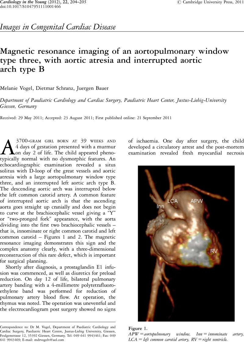
Magnetic resonance imaging of an aortopulmonary window type three, with aortic atresia and interrupted aortic arch type B

Published online by Cambridge University Press:  21 September 2011

Melanie Vogel*
Affiliation:
Department of Paediatric Cardiology and Cardiac Surgery, Paediatric Heart Center, Justus-Liebig-University Giessen, Germany
Dietmar Schranz
Affiliation:
Department of Paediatric Cardiology and Cardiac Surgery, Paediatric Heart Center, Justus-Liebig-University Giessen, Germany
Juergen Bauer
Affiliation:
Department of Paediatric Cardiology and Cardiac Surgery, Paediatric Heart Center, Justus-Liebig-University Giessen, Germany
*
Correspondence to: Dr M. Vogel, Department of Paediatric Cardiology and Cardiac Surgery, Paediatric Heart Centre, Justus-Liebig University, Giessen, Feulgenstrasse 12, 35392 Giessen, Germany. Tel: 049 641 9943461; Fax: 049 641 9943469; E-mail: mdrvogelv@aol.com

Abstract

Image of the first page of this content. For PDF version, please use the ‘Save PDF’ preceeding this image.'
Type
Images in Congenital Cardiac Disease
Copyright
Copyright © Cambridge University Press 2011

Access options

Get access to the full version of this content by using one of the access options below. (Log in options will check for institutional or personal access. Content may require purchase if you do not have access.)

References

1.Yew, G, Coleman, D, Calder, L. Aortic valvar atresia, interrupted aortic arch, and a quadricuspid pulmonary valve: a rare combination. Pediatr Cardiol 2005; 26: 455459.CrossRefGoogle Scholar