


ANCA-associated small vessel vasculitides are systemic diseases characterized by inflammation and necrosis of small-sized blood vessels.Reference Jennette and Falk 1 They are characterized by the presence of antineutrophil cytoplasmic antibodies (ANCAs), which primarily target the kidney, lung, skin and peripheral nervous system.Reference Falk and Jennette 2 In rare cases, they can affect the central nervous system (CNS) and cause cerebrovascular events.Reference Nishino, Rubino, DeRemee, Swanson and Parisi 3 We present a patient with ANCA vasculitis who developed fatal massive bitemporal hemorrhages during the course of immunosuppressive therapy.
A 71-year-old man was referred to our tertiary neurological center due to weight loss (10 kg in two months), fatigue, weakness and persistent fever of unknown origin. The patient did not report previous sinusitis, upper airway infection, arthritis or hematuria. Neurological examination only revealed a right foot drop. Laboratory studies showed inflammatory anemia and elevated inflammatory markers. Screening for infectious diseases was negative. Spine MRI indicated a herniated L5-S1 disc. Brain MRI was normal except for a millimetric left occipital ischemic infarct. Whole-body PET scan was conducted to eliminate a neoplastic etiology, but was also negative. Five days after his arrival, the patient developed new weaknesses and paresthesia in the left leg and right hand. Laboratory studies revealed acute renal failure (creatinine 133 μmol/L, hematuria, proteinuria) and elevated c-ANCA counts (1:320). A second MRI of the brain conducted 10 days after the first showed five new millimetric ischemic foci in the right temporal and left cerebellar regions. In concert with rheumatology, a diagnosis of ANCA vasculitis was made, an immunosuppressive treatment was initiated (high-dose intravenous glucocorticoids), and the patient’s neurological symptoms were stabilized. However, creatinine continued to rise (from 133 to 177 μmol/L in 7 days). A renal biopsy was scheduled to confirm a diagnosis of glomerulonephritis. After five days of induction treatment, tapering oral doses of prednisone were administered. On the first day of prednisone tapering, the patient was found unresponsive in his bed (eyes opened, but unresponsive to simple commands). Head CT showed massive bilateral temporal hemorrhages (see Figure 1). After discussion with the family, the patient was transferred back to his hometown for end-of-life care. Renal biopsy and autopsy could not be performed. Nevertheless, a recent meta-analysis has evaluated the specificity of elevated c-ANCA levels in the serum at 96.3% (CI [94.1-98.5]),Reference Choi, Liu, Merkel, Colditz and Niles 4 which, combined with the high pretest probability of vasculitis (mononeuritis multiplex, acute renal failure, inflammatory anemia, multifocal brain infarcts), made a diagnosis of ANCA vasculitis highly probable even without a gold-standard pathological diagnosis.
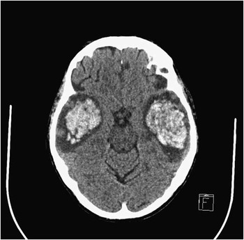
Figure 1 Axial view of a head CT showing massive bitemporal haemorrhages with significant mass effect.
While it is known that ANCA vasculitis may in some instances involve the CNS,Reference Nishino, Rubino, DeRemee, Swanson and Parisi 3 , Reference Choi, Liu, Merkel, Colditz and Niles 4 only a few case reports describe the appearance of cerebral hemorrhages,Reference Akkara Veetil and Schimmer 5 - Reference Han, Rehman, Jayaratne and Carty 10 and even fewer a rapid progression toward massive hemorrhages and death.Reference Akkara Veetil and Schimmer 5 - Reference Sasaki, Hirato, Nakazato, Tanaka and Takeuchi 7 Altogether, our case supports the possibility of cerebral vasculitis, though rare in ANCA-positive patients, causing so massive a cerebral hemorrhage.
ACKNOWLEDGMENTS
We thank the patient’s family for their collaboration. We also thank Audrey Paradis for manuscript formatting. Oral and written consent was obtained from the patient’s family.
Statement of Authorship
David Bergeron drafted the initial version of the manuscript. Dr. Annie Dionne was the treating physician. All authors contributed equally to the writing process and approved the final version of the manuscript.
Disclosures
David Bergeron, Raphaël Marchand and Robert Jr Laforce do not have anything to disclose.
Annie Dionne has the following disclosures: Teva: conducting clinical trials, research grant for the conduct of clinical trials; Novartis: conducting clinical trials, research grant for the conduct of clinical trials.

