


A 45-year-old woman of heritage from the Philippines presented with a 3-week history of intractable hiccups. In the ensuing weeks, she developed intractable vomiting and days before her admission, a change in speech, difficulty swallowing and dizziness were observed. On physical exam, she had oscillopsia, non-fatigable upgaze nystagmus, and mild intention tremor on finger to nose testing bilaterally. Visual exam and fundoscopy were normal. A brain MRI revealed a dorsal medullary periependymal hyperintensity in the area postrema (AP) (Figure 1). The supratentorial white matter was normal. Visual evoked potentials were normal and CSF examination showed normal protein and cell counts. Antinuclear antibodies, infectious, and malignancy screening were negative or normal. Aquaporin-4 antibodies were positive and oligoclonal bands in the CSF were absent.
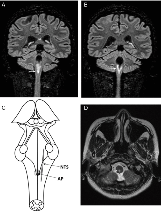
Figure 1: MRI brain coronal T2-FLAIR (A, B) and axial T2-weighted sequences (D) demonstrate an extensive periependymal T2-hyperintense lesion in the dorsal medulla, involving the AP. (C) Schematic representation of the floor of the fourth ventricle, indicating the location of the AP and the NTS.
The central nervous system lesions were thought to be consistent with acute demyelination and the clinical picture that of an area postrema syndrome (APS). She was started on IV methylprednisolone with overall improvement in her hiccups, oscillopsia, and vomiting; however on day 2, she reported new left face numbness. On day 5 of steroids, she complained of saddle anesthesia and bowel dysfunction suggestive of new lesions. A repeat MRI revealed a cord lesion at the C2 level and one in the ventral aspect of the mid thoracic cord (none were longitudinally extensive). Therapy was escalated to plasma exchange (5 sessions). She was diagnosed with aquaporin-4-positive neuromyelitis optica spectrum disorder (NMOSD), according to current criteria. Her neurological symptoms were stabilized, and she was discharged after 17 days.
Hiccups are a common symptom that crosses multiple specialties including gastroenterology, respiratory medicine, and neurology. They are spontaneous myoclonic contractions of the diaphragm and associated muscle groups, thought to be generated by a reflex arc involving afferent, central, and efferent structures of the nervous system.Reference Steger, Schneemann and Fox1 By far, the most common causes of hiccups are gastrointestinal in nature. However, the acute or subacute onset of intractable hiccups has been described in various neurological conditions: medullary strokes, meningitis, epilepsy, intracranial tumors, and demyelinating diseases. The first cases of intractable hiccups associated with demyelinating diseases were reported in 1979.Reference McFarling and Susac2 Three cases of patients with multiple sclerosis (MS) presenting with hiccups were described, and the symptom was thought to reflect disinhibition of a primitive gastrointestinal reflex by demyelinating lesions. Intractable hiccups are now recognized as a classical symptom of APS and characteristic of NMOSD. Other features of APS include intractable nausea and vomiting. Patients have also reported anorexia and significant weight loss.Reference Shosha, Dubey and Palace3
Hiccups are pathophysiologically complex.Reference Steger, Schneemann and Fox1 The afferent impulses are thought to be carried by the vagus, phrenic, or sympathetic nerve fibers. The central areas include the brainstem, the medulla oblongata (respiratory centres, reticular formation), and the upper cervical spinal cord, with special emphasis on the nucleus tractus solitarius (NTS). Many of these structures are, of course, in proximity of the AP, the chemoreceptor-trigger zone for vomiting located in the dorsal surface of the medulla at the caudal end of the fourth ventricle. They are regulated by both dopaminergic and gamma-amino-butyric-acid (GABA-ergic) pathways. The efferent branches involve the innervation of the diaphragm by the phrenic nerve (mid-cervical), innervation of the anterior scalene muscles, innervation of the intercostal muscles (upper thoracic), and finally innervation of the glottis (recurrent branch of the vagus nerve).
In early case series, intractable hiccups and nausea were found to be useful in differentiating between NMOSD and MS. In one such study, 8 out of 47 cases of relapsing NMOSD had intractable hiccups, compared to none in 130 cases of MS.Reference Misu, Fujihara, Nakashima, Sato and Itoyama4 In 75% of patients with intractable hiccups, MRI detected linear medullary lesions involving the pericanal region, the AP, and the NTS. In a more recent series of 258 patients with NMOSD, brainstem signs were observed in 31.4%. Reference Kremer, Mealy and Jacob5 After vomiting, hiccups were the second most common finding (22.3%) and seemed to occur more frequently in non-Caucasians. Some authors have suggested that medullary involvement might herald a more severe disease course. In a case series of 170 patients with NMOSD, 44 were found to have lesions in the medulla oblongata. Reference Wang, Zhang and Zhang6 Lesions in this region were associated with higher rates of hiccups as well as with higher disease severity scores, relapse rates, and longitudinally extensive transverse myelitis.
Whenever possible, the treatment of hiccups should be directed at the underlying cause. In our case, we followed standard management recommendations for an acute demyelinating episode (steroid pulses followed by plasma exchange). In cases where hiccups persist, baclofen and metoclopramide have low-quality evidence for their effectiveness, and there are isolated reports of success with various anticonvulsants. Reference Steger, Schneemann and Fox1 In conclusion, patients who experience intractable hiccups should always prompt a consideration for APS heralding a demyelinating nervous system disease.
Disclosures
The authors have no conflicts of interest to declare.
Statement of Authorship
CRCL drafted the manuscript and produced the figure; DAAS revised the manuscript and refined it for submission; MB revised the manuscript; JJ revised the manuscript and provided intellectual input; JDK revised the manuscript and processed the images; and MY revised the manuscript, provided intellectual input, and follow up information.

