


CLINICIAN'S CAPSULE
What is known about the topic?
A common strategy for managing emergency department patients with low-risk abdominal pain is to arrange for next day outpatient ultrasound.
What did this study ask?
What proportion of outpatient ultrasounds with pathological findings require further evaluation or intervention within 14 days of imaging?
What did this study find?
While the majority of patients did not have ultrasound findings requiring urgent intervention, a significant number (7.7%) did have serious pathology.
Why does this study matter to clinicians?
Clinicians using this deferred management strategy should be aware that a significant proportion of patients may have actionable pathological findings.
INTRODUCTION
Abdominal pain is the most common chief complaint encountered in the emergency department (ED), accounting for approximately 8% of all visits.Reference Graff IV and Robinson1,Reference McCaig and Nawar2 Of these, 25% of patients are discharged from the ED with a diagnosis of undifferentiated abdominal pain.Reference Powers and Guertler3, Reference Kamin, Nowicki, Courtney and Powers4 Studies have shown that most patients discharged with this diagnosis will experience an improvement or resolution of their symptoms without any specific intervention.Reference Kamin, Nowicki, Courtney and Powers4–Reference Weiner, Nagurney, Brown, Sane and Wang6 Despite this, it is common for patients with undifferentiated abdominal pain to have an outpatient ultrasound ordered from the ED to rule out significant pathology. For patients presenting to the ED with abdominal pain on weekends or after hours on weekdays, outpatient ultrasounds with immediate ED reassessment are often arranged. However, a significant proportion of these ultrasounds fail to identify any definitive diagnosis or alter clinical management.Reference Raja, Mortele and Hanson7,Reference Pines8
The objective of this study was to examine the utility of outpatient ultrasounds to assess undifferentiated abdominal pain in patients who are stable enough to be discharged home from the ED and to determine the proportion of those outpatient ultrasounds with pathological findings requiring further evaluation or intervention within 14 days of imaging.
METHODS
This was a retrospective chart review of non-pregnant patients ages 18 to 40 years, presenting to an academic ED (annual census 65,000) with an abdominal complaint for whom the emergency physician arranged an outpatient (next day) abdominal ultrasound. The age range was selected to represent a large proportion of low-risk patients presenting to the ED with abdominal pain who can be safely discharged home with appropriate outpatient follow-up. Pregnant or recently pregnant females and patients admitted to a hospital at the time of the initial ED assessment were excluded. This study protocol was reviewed and approved by the Research Ethics Board at Mount Sinai Hospital in Toronto, Ontario.
Using a computerized, structured data abstraction form, trained research personnel reviewed the medical records from Mount Sinai Hospital and extracted patient data, including patient demographics, ED investigations, follow-up ultrasound interpretation and follow-up interventions. Data were entered directly into a study-specific Microsoft Excel database (Microsoft Corporation, Redmond, WA). Descriptive statistics were summarized using means with standard deviations (SD), medians with interquartile ranges, or frequencies with 95% confidence intervals, where appropriate. Data extraction was done independently and in duplicate for 25% of charts. Interrater agreement was estimated using Cohen's kappa (κ) statistic. Data analysis was performed using SPSS 25.0 (IBM Corporation, Armonk, NY).
RESULTS
Of the 299 included patients, 252 (84.3%) were female and mean (SD) age was 28.4 (6.0) years. Twenty-three (7.7%) patients had ultrasounds requiring intervention within 14 days of imaging. Of these, eight (34.8%) had appendicitis, five (21.7%) had cholecystitis, four (17.4%) had urological pathology, three (13.0%) had gynecological pathology, and three (13.0%) had gastrointestinal diagnoses. The full list of diagnoses is presented in Table 1.
Table 1. Patients requiring medical intervention within 14 days of imaging
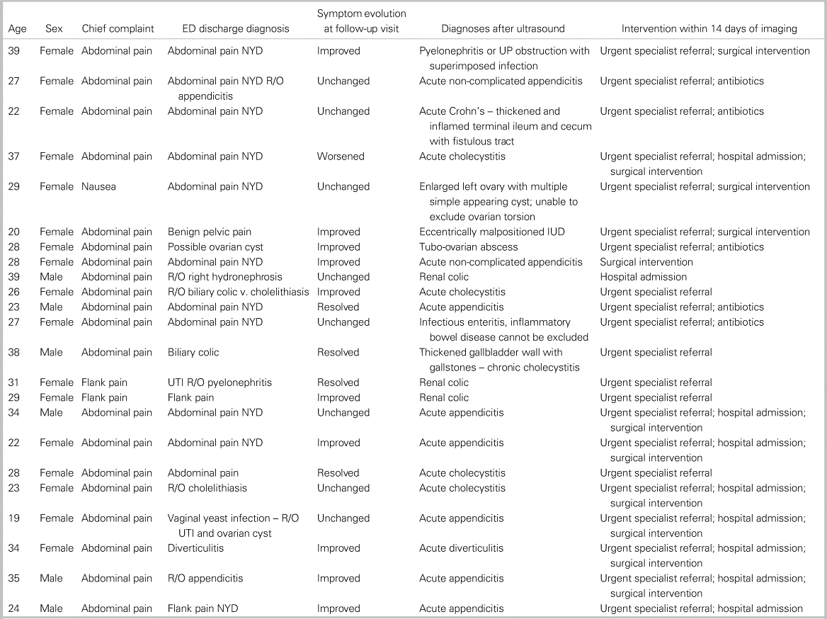
ED = Emergency department; IUD = intrauterine device; NYD = not yet diagnosed; R/O = rule out; UP = ureteropelvic; UTI = urinary tract infection.
Of note, 14 (60.9%) patients requiring intervention within 14 days had symptoms that improved or resolved at the time of the outpatient ultrasound. For the 277 (92.3%) patients not requiring intervention, 117 (42.2%) had improved, 89 (32.1%) were unchanged, 50 (18.1%) had resolved, and 5 (1.8%) had worsened symptoms at the time of the follow-up ultrasound. There were 16 (5.8%) charts that did not contain any information in regard to change in patient-reported symptoms. Of the non-intervention patients, 13 (4.7%) went on to have alternative imaging, including magnetic resonance imaging, computed tomography, and sonohysterogram.
DISCUSSION
The objective of this study was to examine a strategy of deferred (scheduled next day) ultrasound investigations and ED follow-up for patients with low-risk abdominal pain, and to determine the proportion of those patients with important findings requiring treatment or intervention within 14 days. While the majority of patients who managed using this strategy did not have ultrasound findings requiring urgent intervention or treatment, a significant number (7.7%) did have serious pathology. This study suggests that this strategy may be a reasonable one, especially after hours when access to ultrasound is limited for this particular patient population. However, our findings suggest that clinicians opting to use this deferred management strategy need to be aware that a significant proportion of patients may in fact have actionable pathological findings.
A second finding from our study that could impact patient management is that 60.9% patients requiring intervention within 14 days had symptoms that improved or resolved at the time of the outpatient ultrasound. The significance of this finding is not clear; it is possible that the natural history of some cases of appendicitis and cholecystitis is to simply resolve on their own, but it seems unlikely this would account for the majority of patients with resolved abdominal pain in our study. Future studies should attempt to elucidate which, if any, clinical features from the initial ED presentation are predictive of significant pathology on outpatient ultrasound. Additionally, as emergency provider point-of-care ultrasound skills continue to advance, it is possible that fewer patients with significant abnormalities will be sent home for next day imaging. At the time of this study, over 95% of the physicians working in this ED were Canadian Emergency Ultrasound Independent Practitioner certified for point-of-care ultrasound.
Unnecessary diagnostic imaging contributes to rising healthcare costs and carries the potential for harm and inconvenience to patients with incidental findings, leading to further investigation and, occasionally, unwarranted procedures. These additional investigations are costly at both the system and patient levels. At the system level, such testing uses scarce and expensive healthcare resources to chase what often turns out to be completely incidental findings. Additionally, the implications of a next-day compared with same-day ultrasound strategy on patient flow and ED wait times are unknown. At the patient level, in addition to the anxiety associated with abnormal results, follow-up investigations are frequently invasive, have direct time and transportation associated costs, and expose patients to additional radiation, contrast dye, and infection risks that may contribute to patient morbidity.
Limitations
This study is not without limitations. Due to the retrospective nature of this study, we can only report what was documented in the patient record. This was a single-centre study, and while clinical management strategies may be similar at other institutions, our findings may not be generalizable to other settings. It is possible that a patient may have re-presented to another hospital in the same city and received treatment or had an intervention not captured in this study. It is possible that some of the observed variation in the collected data were influenced by error or bias from the data abstractor, who was not blinded to the objective of this descriptive study. Additionally, our study could not capture the thought process and patient-physician exchanges that were involved in the imaging decision. It is possible that ED management was dictated by patient preference, point-of-care ultrasound findings, or other information not documented in the chart.
CONCLUSIONS
The large majority of patients with abdominal pain discharged from the ED with planned next-day ultrasound had findings that were normal or did not require any additional management. However, 7.7% of these patients had pathological findings that required intervention within 14 days. Interestingly, many of those patients had pain that had resolved or improved by the next day. Next-day ultrasound imaging remains a good way of identifying patients with serious pathology not appreciated at the time of their ED visit, and may be better at identifying patients with significant problems who may have had delayed presentation if simply instructed to return to the ED if their symptoms had not improved in 24 to 48 hours.
Author contributions
The authors all stand behind the conclusions of this manuscript, agree to be accountable for all aspects of the work, and support its publication. All authors contributed to the study conception and designed the protocol. All authors contributed to the manuscript preparation and have given approval for its submission.
Competing interests
None declared.

