Skip to main content Accessibility help
×
Hostname: page-component-76fb5796d-vvkck Total loading time: 0 Render date: 2024-04-25T07:59:33.447Z Has data issue: false hasContentIssue false

7 - Magnetic Nanoparticles for Magnetic Resonance Imaging Contrast Agents

Published online by Cambridge University Press:  10 February 2019

Nicholas J. Darton
Affiliation:
Arecor Limited
Adrian Ionescu
Affiliation:
University of Cambridge
Justin Llandro
Affiliation:
Tohoku University, Japan
Get access

Summary

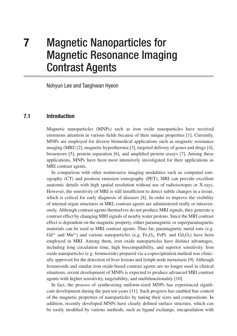
Image of the first page of this content. For PDF version, please use the ‘Save PDF’ preceeding this image.'
Type
Chapter
Information
Publisher: Cambridge University Press
Print publication year: 2019

Access options

Get access to the full version of this content by using one of the access options below. (Log in options will check for institutional or personal access. Content may require purchase if you do not have access.)

References

Lu, A-H., Salabas, E. L., and Schueth, F. Magnetic nanoparticles: Synthesis, protection, functionalization, and application. Angew. Chem. Int. Ed., 46:8 (2007), 1222–44.Google Scholar
Laurent, S., Forge, D., and Port, M., et al., Magnetic iron oxide nanoparticles: Synthesis, stabilization, vectorization, physicochemical characterizations, and biological applications. Chem. Rev., 108:6 (2008), 2064–110.Google Scholar
Fortin, J-P., Wilhelm, C., Servais, J., Menager, C., Bacri, J-C., and Gazeau, F. Size-sorted anionic iron oxide nanomagnets as colloidal mediators for magnetic hyperthermia. J. Am. Chem. Soc., 129:9 (2007), 2628–35.CrossRefGoogle ScholarPubMed
Cheng, K., Peng, S., Xu, C., and Sun, S. Porous hollow Fe3O4 nanoparticles for targeted delivery and controlled release of cisplatin. J. Am. Chem. Soc., 131:30 (2009), 10637–44.Google Scholar
Lee, H., Yoon, T-J., Figueiredo, J-L., Swirski, F. K., and Weissleder, R. Rapid detection and profiling of cancer cells in fine-needle aspirates. Proc. Natl. Acad. Sci. USA., 106:30 (2009), 12459–64.Google ScholarPubMed
Lee, I. S., Lee, N., Park, J., et al., Ni/NiO core/shell nanoparticles for selective binding and magnetic separation of histidine-tagged proteins. J. Am. Chem. Soc., 128:33 (2006), 10658–9.Google Scholar
Rosi, N. L. and Mirkin, C. A. Nanostructures in biodiagnostics. Chem. Rev. 105:4 (2005), 1547–62.CrossRefGoogle ScholarPubMed
Willmann, J. K., Van Bruggen, N., Dinkelborg, L. M., and Gambhir, S. S. Molecular imaging in drug development. Nat. Rev. Drug Discov., 7:7 (2008), 591607.Google Scholar
Harisinghani, M. G., Barentsz, J., Hahn, P. F., et al., Noninvasive detection of clinically occult lymph-node metastases in prostate cancer. N. Engl. J. Med., 348:25 (2003), 2491–9.Google Scholar
Lee, N. and Hyeon, T. Designed synthesis of uniformly sized iron oxide nanoparticles for efficient magnetic resonance imaging contrast agents. Chem. Soc. Rev., 41:7 (2012), 2575–89.Google Scholar
Hao, R., Xing, R., Xu, Z., Hou, Y., Gao, S., and Sun, S. Synthesis, functionalization, and biomedical applications of multifunctional magnetic nanoparticles. Adv. Mater., 22:25 (2010), 2729–42.CrossRefGoogle ScholarPubMed
Xie, J., Liu, G., Eden, H. S., Ai, H., and Chen, X. Surface-engineered magnetic nanoparticle platforms for cancer imaging and therapy. Acc. Chem. Res., 44:10 (2011), 883–92.Google Scholar
Aime, S., Castelli, D. D., Crich, S. G., Gianolio, E., and Terreno, E. Pushing the sensitivity envelope of lanthanide-based magnetic resonance imaging (MRI) contrast agents for molecular imaging applications. Acc. Chem. Res., 42:7 (2009), 822–31.Google Scholar
Sieber, M. A., Steger-Hartmann, T., Lengsfeld, P., and Pietsch, H. Gadolinium-based contrast agents and nsf: evidence from animal experience. J. Magn. Reson. Imaging, 30:6 (2009), 1268–76.Google Scholar
Kim, D., Lee, N., Park, M., Kim, B. H., An, K., and Hyeon, T. Synthesis of uniform ferrimagnetic magnetite nanocubes. J. Am. Chem. Soc., 131:2 (2009), 454–5.Google ScholarPubMed
Morales, M. P., Veintemillas-Verdaguer, S., Montero, M. I., et al., Surface and internal spin canting in γ-Fe2O3 nanoparticles. Chem. Mater., 11:11 (1999), 3058–64.Google Scholar
Kim, B. H., Lee, N., Kim, H., et al., Large-scale synthesis of uniform and extremely small-sized iron oxide nanoparticles for high-resolution T1 magnetic resonance imaging contrast agents. J. Am. Chem. Soc., 133:32 (2011), 12624–31.CrossRefGoogle ScholarPubMed
Park, J., An, K., Hwang, Y. S., et al., Ultra-large-scale syntheses of monodisperse nanocrystals. Nat. Mater., 3:12 (2004), 891–5.CrossRefGoogle ScholarPubMed
Brooks, R. A., Moiny, F., and Gillis, P. On T2-shortening by weakly magnetized particles: The chemical exchange model. Magn. Reson. Med., 45:6 (2001), 1014–20.Google Scholar
Gillis, P., Moiny, F., and Brooks, R. A. On T2-shortening by strongly magnetized spheres: A partial refocusing model. Magn. Reson. Med., 47:2 (2002), 257–63.Google Scholar
Jun, Y. W., Huh, Y-M., Choi, J. S., et al., Nanoscale size effect of magnetic nanocrystals and their utilization for cancer diagnosis via magnetic resonance imaging. J. Am. Chem. Soc., 127:16 (2005), 5732–3.CrossRefGoogle ScholarPubMed
Lee, N., Kim, H., Choi, S. H., et al., Magnetosome-like ferrimagnetic iron oxide nanocubes for highly sensitive MRI of single cells and transplanted pancreatic islets. Proc. Natl. Acad. Sci. USA., 108:7 (2011), 2662–7.Google Scholar
Li, W., Tutton, S., Vu, A. T., et al., First-pass contrast-enhanced magnetic resonance angiography in humans using ferumoxytol, a novel ultrasmall superparamagnetic iron oxide (USPIO)-based blood pool agent. J. Magn. Reson. Imaging, 21:1 (2005), 4652.CrossRefGoogle ScholarPubMed
Lee, J-H., Huh, Y-M., Jun, Y-W., et al., Artificially engineered magnetic nanoparticles for ultra-sensitive molecular imaging. Nat. Med., 13:1 (2007), 95–9.CrossRefGoogle ScholarPubMed
Jang, J-T., Nah, H., Lee, J-H., Moon, S. H., Kim, M. G., and Cheon, J. Critical enhancements of MRI contrast and hyperthermic effects by dopant-controlled magnetic nanoparticles. Angew. Chem. Int. Ed., 48:7 (2009), 1234–8.Google Scholar
Chaubey, G. S., Barcena, C., Poudyal, N., et al., Synthesis and stabilization of FeCo nanoparticles. J. Am. Chem. Soc., 129:23 (2007), 7214–5.Google Scholar
Seo, W. S., Lee, J. H., Sun, X., et al., FeCo/graphitic-shell nanocrystals as advanced magnetic-resonance-imaging and near-infrared agents. Nat. Mater., 5:12 (2006), 971–6.Google Scholar
Lee, J. H., Sherlock, S. P., Terashima, M., et al., High-contrast in vivo visualization of microvessels using novel FeCo/GC magnetic nanocrystals. Magn. Reson. Med., 62:6 (2009), 1497–509.CrossRefGoogle ScholarPubMed
Cheong, S., Ferguson, P., Feindel, K. W., et al., Simple synthesis and functionalization of iron nanoparticles for magnetic resonance imaging. Angew. Chem. Int. Ed., 50:18 (2011), 4206–9.Google Scholar
Hyeon, T., Lee, S. S., Park, J., Chung, Y., and Na, H. B. Synthesis of highly crystalline and monodisperse maghemite nanocrystallites without a size-selection process. J. Am. Chem. Soc., 123:51 (2001), 12798–801.CrossRefGoogle ScholarPubMed
Peng, S., Wang, C., Xie, J., and Sun, S. Synthesis and stabilization of monodisperse Fe nanoparticles. J. Am. Chem. Soc., 128:33 (2006), 10676–7.Google Scholar
Lee, H., Yoon, T-J., and Weissleder, R. Ultrasensitive detection of bacteria using core-shell nanoparticles and an NMR-filter system. Angew. Chem. Int. Ed., 48:31 (2009), 5657–60.Google Scholar
Yoon, T-J., Lee, H., Shao, H., and Weissleder, R. Highly magnetic core-shell nanoparticles with a unique magnetization mechanism. Angew. Chem. Int. Ed., 50:20 (2011), 4663–6.Google Scholar
Yoon, T-J., Lee, H., Shao, H., Hilderbrand, S. A., and Weissleder, R. Multicore assemblies potentiate magnetic properties of biomagnetic nanoparticles. Adv. Mater., 23:41 (2011), 47934797.Google Scholar
Perez, J. M., Josephson, L., ‘Loughlin, T. O’, Högemann, D., and Weissleder, R. Magnetic relaxation switches capable of sensing molecular interactions. Nat. Biotechnol., 20:8 (2002), 816–20.CrossRefGoogle ScholarPubMed
Brooks, R. A. T2-shortening by strongly magnetized spheres: A chemical exchange model. Magn. Reson. Med. 47:2 (2002), 388–91.CrossRefGoogle Scholar
Tromsdorf, U. I., Bigall, N. C., Kaul, M. G., et al., Size and surface effects on the MRI relaxivity of manganese ferrite nanoparticle contrast agents. Nano Lett., 7:8 (2007), 2422–7.Google Scholar
Bowen, C. V., Zhang, X., Saab, G., Gareau, P. J., and Rutt, B. K. Application of the static dephasing regime theory to superparamagnetic iron-oxide loaded cells. Magn. Reson. Med., 48:1 (2002), 5261.Google Scholar
Lee, J. E., Lee, N., Kim, T., Kim, J., and Hyeon, T. Multifunctional mesoporous silica nanocomposite nanoparticles for theranostic applications. Acc. Chem. Res., 44:10 (2011), 893902.CrossRefGoogle ScholarPubMed
Lee, J-H., Jun, Y-W., Yeon, S-I., Shin, J-S., and Cheon, J. Dual-mode nanoparticle probes for high-performance magnetic resonance and fluorescence imaging of neuroblastoma. Angew. Chem. Int. Ed., 45:48 (2006), 8160–2.Google Scholar
Lee, J. E., Lee, N., Kim, H., et al., Uniform mesoporous dye-doped silica nanoparticles decorated with multiple magnetite nanocrystals for simultaneous enhanced magnetic resonance imaging, fluorescence imaging, and drug delivery. J. Am. Chem. Soc., 132:2 (2010), 552–7.Google ScholarPubMed
Tong, S., Hou, S., Zheng, Z., Zhou, J., and Bao, G. Coating optimization of superparamagnetic iron oxide nanoparticles for high T2 relaxivity. Nano Lett., 10:11 (2010), 4607–13.Google Scholar
Granot, D. and Shapiro, E. M. Release activation of iron oxide nanoparticles: (REACTION) a novel environmentally sensitive MRI paradigm. Magn. Reson. Med., 65:5 (2011), 1253–9.Google Scholar
Kaittanis, C., Santra, S., Santiesteban, O. J., Henderson, T. J., and Perez, J. M. The assembly state between magnetic nanosensors and their targets orchestrates their magnetic relaxation response. J. Am. Chem. Soc., 133:10 (2011), 3668–76.Google Scholar
Dubertret, B., Skourides, P., Norris, D. J., Noireaux, V., Brivanlou, A. H., and Libchaber, A. In vivo imaging of quantum dots encapsulated in phospholipid micelles. Science, 298:5599 (2002), 1759–62.Google Scholar
Ling, D., Park, W., Park, Y. I., et al., Multiple-interaction ligands inspired by mussel adhesive protein: synthesis of highly stable and biocompatible nanoparticles. Angew. Chem. Int. Ed., 50:48 (2011), 11360–5.Google Scholar
Perazella, M. A. Current status of gadolinium toxicity in patients with kidney disease. Clin. J. Am. Soc. Nephrol., 4:2 (2009), 461–9.Google Scholar
Choi, H. S., Liu, W., Misra, P., et al., Renal clearance of quantum dots. Nat. Biotechnol., 25:10 (2007), 1165–70.Google Scholar
Arbab, A. S., Wilson, L. B., Ashari, P., Jordan, E. K., Lewis, B. K., and Frank, J. A. A model of lysosomal metabolism of dextran coated superparamagnetic iron oxide (SPIO) nanoparticles: Implications for cellular magnetic resonance imaging. NMR Biomed., 18:6 (2005), 383–9.Google Scholar
Gao, J., Liang, G., Zhang, B., Kuang, Y., Zhang, X., and Xu, B. FePt@CoS2 yolk-shell nanocrystals as a potent agent to kill HeLa cells. J. Am. Chem. Soc., 129:5 (2007), 1428–33.CrossRefGoogle ScholarPubMed
Bourrinet, P., Bengele, H. H., Bonnemain, B., et al., Preclinical safety and pharmacokinetic profile of ferumoxtran-10, an ultrasmall superparamagnetic iron oxide magnetic resonance contrast agent. Invest. Radiol., 41:3 (2006), 313–24.CrossRefGoogle ScholarPubMed

Save book to Kindle

To save this book to your Kindle, first ensure coreplatform@cambridge.org is added to your Approved Personal Document E-mail List under your Personal Document Settings on the Manage Your Content and Devices page of your Amazon account. Then enter the ‘name’ part of your Kindle email address below. Find out more about saving to your Kindle.

Note you can select to save to either the @free.kindle.com or @kindle.com variations. ‘@free.kindle.com’ emails are free but can only be saved to your device when it is connected to wi-fi. ‘@kindle.com’ emails can be delivered even when you are not connected to wi-fi, but note that service fees apply.

Find out more about the Kindle Personal Document Service.

Available formats
×

Save book to Dropbox

To save content items to your account, please confirm that you agree to abide by our usage policies. If this is the first time you use this feature, you will be asked to authorise Cambridge Core to connect with your account. Find out more about saving content to Dropbox.

Available formats
×

Save book to Google Drive

To save content items to your account, please confirm that you agree to abide by our usage policies. If this is the first time you use this feature, you will be asked to authorise Cambridge Core to connect with your account. Find out more about saving content to Google Drive.

Available formats
×