Skip to main content Accessibility help
×
Hostname: page-component-76fb5796d-45l2p Total loading time: 0 Render date: 2024-04-25T17:25:13.568Z Has data issue: false hasContentIssue false

19 - High Resolution Imaging in the Graphene Liquid Cell

from Part III - Prospects

Published online by Cambridge University Press:  22 December 2016

Frances M. Ross
Affiliation:
IBM T. J. Watson Research Center, New York
Get access

Summary

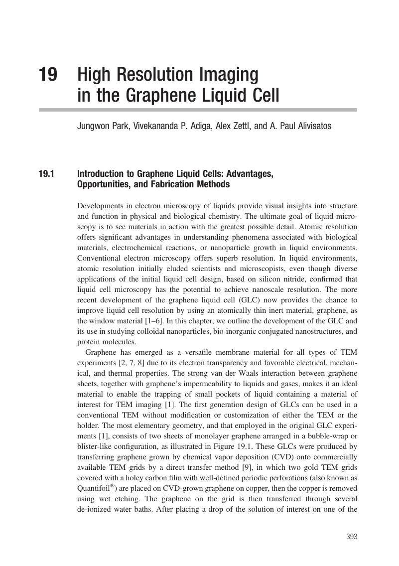
Image of the first page of this content. For PDF version, please use the ‘Save PDF’ preceeding this image.'
Type
Chapter
Information
Publisher: Cambridge University Press
Print publication year: 2016

Access options

Get access to the full version of this content by using one of the access options below. (Log in options will check for institutional or personal access. Content may require purchase if you do not have access.)

References

Yuk, J. M., Park, J., Ercius, P. et al., High-resolution EM of colloidal nanocrystal growth using graphene liquid cells. Science, 336 (2012), 6164.Google Scholar
Chen, Q., Smith, J. M., Park, J. et al., 3D motion of DNA-Au nanoconjugates in graphene liquid cell electron microscopy. Nano Lett., 13 (2013), 45564561.Google Scholar
Wang, C., Qiao, Q., Shokuhfar, T. and Klie, R. F., High-resolution electron microscopy and spectroscopy of ferritin in biocompatible graphene liquid cells and graphene sandwiches. Adv. Mater., 26 (2014), 34103414.CrossRefGoogle ScholarPubMed
De Clercq, A., Dachraoui, W., Margeat, O. et al., Growth of Pt–Pd nanoparticles studied in situ by HRTEM in a liquid cell. J. Phys. Chem. Lett., 5 (2014), 21262130.Google Scholar
Mohanty, N., Fahrenholtz, M., Nagaraja, A., Boyle, D. and Berry, V., Impermeable graphenic encasement of bacteria. Nano Lett., 11 (2011), 12701275.Google Scholar
Ericius, P., Kim, K., Zettl, A. et al., In-situ observations of Pt nanoparticle growth at atomic resolution using graphene liquid cells and Cc correction. Microsc. Microanal., 18 (2012), 10961097.CrossRefGoogle Scholar
Nair, R. R., Blake, P., Blake, J. R. et al., Graphene as a transparent conductive support for studying biological molecules by transmission electron microscopy. Appl. Phys. Lett., 97 (2010), 153102.CrossRefGoogle Scholar
Lee, Z., Jeon, K., Dato, A. and Erni, R., Direct imaging of soft–hard interfaces enabled by graphene. Nano Lett., 9 (2009), 33653369.Google Scholar
Regan, W., Alem, N., Alemán, B. et al., A direct transfer of layer-area graphene. Appl. Phys. Lett., 96 (2010), 113102.Google Scholar
Evans, J. E., Jungjohann, K. L., Browning, N. D. and Arslan, I., Controlled growth of nanoparticles from solution with in situ liquid transmission electron microscopy. Nano Lett., 11 (2011), 28092813.Google Scholar
Zheng, H., Claridge, S. A, Minor, A. M., Alivisatos, A. P., and Dahmen, U., Nanocrystal diffusion in a liquid thin film observed by in situ transmission electron microscopy. Nano Lett., 9 (2009), 24602465.Google Scholar
Woehl, T. J., Evans, J. E., Arslan, I., Ristenpart, W. D. and Browning, N. D., Direct in situ determination of the mechanisms controlling nanoparticle nucleation and growth. ACS Nano, 6 (2012), 85998610.CrossRefGoogle ScholarPubMed
Mirsaidov, U. M., Zheng, H., Casana, Y. and Matsudaira, P., Imaging protein structure in water at 2.7 nm resolution by transmission electron microscopy. Biophys. J., 102 (2012), L15L17.CrossRefGoogle Scholar
Zheng, H., Mirsaidov, U. M., Wang, L.-W. and Matsudaira, P., Electron beam manipulation of nanoparticles. Nano Lett., 12 (2012), 56445648.CrossRefGoogle ScholarPubMed
Proetto, M. T., Rush, A. M., Chien, M.-P. et al., Dynamics of soft nanomaterials captured by transmission electron microscopy in liquid water. J. Am. Chem. Soc., 136 (2014), 11621165.Google Scholar
Jungjohann, K. L., Bliznakov, S., Sutter, P. W., Stach, E. A and Sutter, E. A., In situ liquid cell electron microscopy of the solution growth of Au-Pd core-shell nanostructures. Nano Lett., 13 (2013), 29642970.Google Scholar
Mirsaidov, U. M., Zheng, H., Bhattacharya, D., Casana, Y. and Matsudaira, P., Direct observation of stick-slip movements of water nanodroplets induced by an electron beam. Proc. Natl. Acad. Sci. USA, 109 (2012), 71877190.Google Scholar
Parent, L. R., Robinson, D. B., Cappillino, P. J. et al., In situ observation of directed nanoparticle aggregation during the synthesis of ordered nanoporous metal in soft templates. Chem. Mater., 26 (2014), 14261433.Google Scholar
Zheng, H., Smith, R. K., Jun, Y.-W. et al., Observation of single colloidal platinum nanocrystal growth trajectories. Science, 324 (2009), 13091312.Google Scholar
Liao, H.-G., Cui, L., Whitelam, S. and Zheng, H., Real-time imaging of Pt3Fe nanorod growth in solution. Science, 336 (2012), 10111014.Google Scholar
Park, J., Zheng, H., Lee, W. C. et al., Direct observation of nanoparticle superlattice formation by using liquid cell transmission electron microscopy. ACS Nano, 6 (2012), 20782085.Google Scholar
Grogan, J. M., Schneider, N. M., Ross, F. M. and Bau, H. H., Bubble and pattern formation in liquid induced by an electron beam. Nano Lett., 14 (2014), 359364.CrossRefGoogle ScholarPubMed
Schneider, N. M., Norton, M. M., Mendel, B. J. et al., Electron–water interactions and implications for liquid cell electron microscopy. J. Phys. Chem. C, 118 (2014), 2237322382.Google Scholar
Adiga, V. P., Dunn, G. D., Alivisatos, A. P. and Zettl, A., Liquid flow cells having graphene on nitride for microscopy. US Patent Application No. US 20160042912 A1.Google Scholar
Holtz, M. E., Yu, Y., Gao, J., Abruña, H. D. and Muller, D. A., In situ electron energy-loss spectroscopy in liquids. Microsc. Microanal., 19 (2013), 10271035.Google Scholar
Polte, J., Erler, R. and Thu, A. F. et al., Nucleation and growth of gold nanoparticles studied via in situ small angle X-ray scattering at millisecond time resolution. ACS Nano, 4 (2010), 10761082.Google Scholar
Harada, M. and Katagiri, E., Mechanism of silver particle formation during photoreduction using in situ time-resolved SAXS analysis. Langmuir, 26 (2010), 1789617905.CrossRefGoogle ScholarPubMed
Polte, J., Ahner, T. T., Delissen, F. et al., Mechanism of gold nanoparticle formation in the classical citrate synthesis method derived from coupled in situ XANES and SAXS evaluation. J. Am. Chem. Soc., 132 (2010), 12961301.Google Scholar
Lu, X., Rycenga, M., Skrabalak, S. E., Wiley, B. and Xia, Y., Chemical synthesis of novel plasmonic nanoparticles. Annu. Rev. Phys. Chem., 60 (2009), 167192.Google Scholar
Schapotschnikow, P., Pool, R. and Vlugt, T. J. H., Molecular simulations of interacting nanocrystals. Nano Lett., 8 (2008), 29302934.Google Scholar

Save book to Kindle

To save this book to your Kindle, first ensure coreplatform@cambridge.org is added to your Approved Personal Document E-mail List under your Personal Document Settings on the Manage Your Content and Devices page of your Amazon account. Then enter the ‘name’ part of your Kindle email address below. Find out more about saving to your Kindle.

Note you can select to save to either the @free.kindle.com or @kindle.com variations. ‘@free.kindle.com’ emails are free but can only be saved to your device when it is connected to wi-fi. ‘@kindle.com’ emails can be delivered even when you are not connected to wi-fi, but note that service fees apply.

Find out more about the Kindle Personal Document Service.

Available formats
×

Save book to Dropbox

To save content items to your account, please confirm that you agree to abide by our usage policies. If this is the first time you use this feature, you will be asked to authorise Cambridge Core to connect with your account. Find out more about saving content to Dropbox.

Available formats
×

Save book to Google Drive

To save content items to your account, please confirm that you agree to abide by our usage policies. If this is the first time you use this feature, you will be asked to authorise Cambridge Core to connect with your account. Find out more about saving content to Google Drive.

Available formats
×