Skip to main content Accessibility help
×
Hostname: page-component-8448b6f56d-qsmjn Total loading time: 0 Render date: 2024-04-23T22:10:43.506Z Has data issue: false hasContentIssue false

10 - Micro- and nanotools to probe cancer cell mechanics and mechanobiology

from Part I - Micro-nano techniques in cell mechanobiology

Published online by Cambridge University Press:  05 November 2015

Yu Sun
Affiliation:
University of Toronto
Deok-Ho Kim
Affiliation:
University of Washington
Craig A. Simmons
Affiliation:
University of Toronto
Get access

Summary

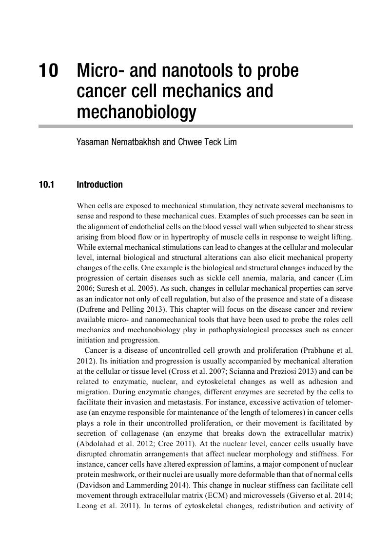
Image of the first page of this content. For PDF version, please use the ‘Save PDF’ preceeding this image.'
Type
Chapter
Information
Integrative Mechanobiology
Micro- and Nano- Techniques in Cell Mechanobiology
, pp. 169 - 185
Publisher: Cambridge University Press
Print publication year: 2015

Access options

Get access to the full version of this content by using one of the access options below. (Log in options will check for institutional or personal access. Content may require purchase if you do not have access.)

References

Abdolahad, M., Sanaee, Z., Janmaleki, M., et al. (2012). “Vertically aligned multiwall-carbon nanotubes to preferentially entrap highly metastatic cancerous cells.” Carbon 50(5): 20102017.CrossRefGoogle Scholar
Adamo, A., Sharei, A., Adamo, L., et al. (2012). “Microfluidics-based assessment of cell deformability.” Anal Chem 84(15): 64386443.CrossRefGoogle ScholarPubMed
Ashkin, A. and Dziedzic, J. M.. (1987). “Optical trapping and manipulation of viruses and bacteria.” Science 235(4795): 15171520.CrossRefGoogle ScholarPubMed
Babahosseini, H., Srinivasaraghavan, V. and Agah, M.. (2012). “Microfluidic chip bio-sensor for detection of cancer cells.” Sensors. Taipei, IEEE 14, 28–31 Oct. 2012.Google Scholar
Badique, F., Stamov, D. R., Davidson, P. M., et al. (2013). “Directing nuclear deformation on micropillared surfaces by substrate geometry and cytoskeleton organization.” Biomaterials 34(12): 29913001.CrossRefGoogle ScholarPubMed
Beebe, D. J., Mensing, G. A. and Walker, G. M.. (2002). “Physics and applications of microfluidics in biology.” Annu Rev Biomed Eng 4: 261286.CrossRefGoogle Scholar
Bellini, N., Bragheri, F., Cristiani, I., et al. (2012). “Validation and perspectives of a femtosecond laser fabricated monolithic optical stretcher.” Biomed Opt Express 3(10): 26582668.CrossRefGoogle ScholarPubMed
Binnig, G., Quate, C. F. and Gerber, C.. (1986). “Atomic force microscope.” Phys Rev Lett 56(9): 930933.CrossRefGoogle ScholarPubMed
Byun, S., Son, S., Amodei, D., et al. (2013). “Characterizing deformability and surface friction of cancer cells.” Proc Natl Acad Sci USA 110(19): 6.CrossRefGoogle ScholarPubMed
Chen, C. L., Mahalingam, D., Osmulski, P., et al. (2013). “Single-cell analysis of circulating tumor cells identifies cumulative expression patterns of EMT-related genes in metastatic prostate cancer.” Prostate 73(8): 813826.CrossRefGoogle ScholarPubMed
Cheng, G., Tse, J., Jain, R. K., et al. (2009). “Micro-environmental mechanical stress controls tumor spheroid size and morphology by suppressing proliferation and inducing apoptosis in cancer cells.” PLoS One 4(2): e4632.CrossRefGoogle ScholarPubMed
Cheung, L. S., Zheng, X., Stopa, A., et al. (2009). “Detachment of captured cancer cells under flow acceleration in a bio-functionalized microchannel.” Lab Chip 9(12): 17211731.CrossRefGoogle Scholar
Cree, I. A. (2011). “Principles of cancer cell culture.” Methods Mol Biol 731: 1326.CrossRefGoogle ScholarPubMed
Crick, F. H. C., and Hughes, A. F. W. (1950). “The physical properties of cytoplasm: a study by means of the magnetic particle method.” Experimental Cell Research 1(1): 44.Google Scholar
Cross, S. E., Jin, Y. S., Rao, J., et al. (2007). “Nanomechanical analysis of cells from cancer patients.” Nat Nanotechnol 2(12): 780783.CrossRefGoogle ScholarPubMed
Darling, E. M., Zauscher, S., Block, J. A., et al. (2007). “A thin-layer model for viscoelastic, stress-relaxation testing of cells using atomic force microscopy: do cell properties reflect metastatic potential?Biophys J 92(5): 17841791.CrossRefGoogle ScholarPubMed
Davidson, P. M. and Lammerding, J.. (2014). “Broken nuclei–lamins, nuclear mechanics, and disease.” Trends Cell Biol 24(4): 247256.CrossRefGoogle ScholarPubMed
Dudani, J. S., Gossett, D. R., Tse, H. T., et al. (2013). “Pinched-flow hydrodynamic stretching of single-cells.” Lab Chip 13(18): 37283734.CrossRefGoogle ScholarPubMed
Dufrene, Y. F. and Pelling, A. E.. (2013). “Force nanoscopy of cell mechanics and cell adhesion.” Nanoscale 5(10): 40944104.CrossRefGoogle ScholarPubMed
Faria, E. C., Ma, N., Gazi, E., et al. (2008). “Measurement of elastic properties of prostate cancer cells using AFM.” Analyst 133(11): 14981500.CrossRefGoogle ScholarPubMed
Faustino, V., Pinho, D., Yaginuma, T., et al. (2014). “Extensional flow-based microfluidic device: deformability assessment of red blood cells in contact with tumor cells.” BioChip Journal 8(1): 4247.CrossRefGoogle Scholar
Fu, C., Han, C., Cheng, C., et al. (2012). “Bio-mechanical properties of human renal cancer cells probed by magneto-optical tweezers.” Sensors. Taipei, IEEE, 14, 28–31 Oct. 2012.Google Scholar
Fu, Y., Vandongen, A. M. J., Bourouina, T., et al. (2012). “A study of cancer cell metastasis using microfluidic transmigration device.” MEMS. Paris, IEEE: 773776, 29 Jan. 2012–2 Feb. 2012.Google Scholar
Giverso, C., Grillo, A. and Preziosi, L.. (2014). “Influence of nucleus deformability on cell entry into cylindrical structures.” Biomech Model Mechanobiol 13(3): 481502.CrossRefGoogle ScholarPubMed
Gossett, D. R., Tse, H. T., Lee, S. A., et al. (2012). “Hydrodynamic stretching of single cells for large population mechanical phenotyping.” Proc Natl Acad Sci USA 109(20): 76307635.CrossRefGoogle ScholarPubMed
Guck, J., Ananthakrishnan, R., Mahmood, H., et al. (2001). “The optical stretcher: a novel laser tool to micromanipulate cells.” Biophys J 81(2): 767784.CrossRefGoogle ScholarPubMed
Guck, J., Schinkinger, S., Lincoln, B., et al. (2005). “Optical deformability as an inherent cell marker for testing malignant transformation and metastatic competence.” Biophys J 88(5): 36893698.CrossRefGoogle ScholarPubMed
Guo, Q., Park, S. and Ma, H.. (2012). “Microfluidic micropipette aspiration for measuring the deformability of single cells.” Lab Chip 12(15): 26872695.CrossRefGoogle ScholarPubMed
Hou, H. W., Li, Q. S., Lee, G. Y., et al. (2009). “Deformability study of breast cancer cells using microfluidics.” Biomed Microdevices 11(3): 557564.CrossRefGoogle ScholarPubMed
Jaeger, A. A., Das, C. K., Morgan, N. Y., et al. (2013). “Microfabricated polymeric vessel mimetics for 3-D cancer cell culture.” Biomaterials 34(33): 83018313.CrossRefGoogle ScholarPubMed
Ketene, A. N., Schmelz, E. M., Roberts, P. C., et al. (2012). “The effects of cancer progression on the viscoelasticity of ovarian cell cytoskeleton structures.” Nanomedicine 8(1): 93102.CrossRefGoogle ScholarPubMed
Kirmizis, D. and Logothetidis, S.. (2010). “Atomic force microscopy probing in the measurement of cell mechanics.” Int J Nanomedicine 5: 137145.CrossRefGoogle ScholarPubMed
Kittur, H., Weaver, W. and Di Carlo, D.. (2014). “Well-plate mechanical confinement platform for studies of mechanical mutagenesis.” Biomed Microdevices 16(3): 439447.CrossRefGoogle ScholarPubMed
Kozissnik, B. and Dobson, J.. (2013). “Biomedical applications of mesoscale magnetic particles.” MRS Bulletin 38(11): 927932.CrossRefGoogle Scholar
Krause, M., Te Riet, J. and Wolf, K.. (2013). “Probing the compressibility of tumor cell nuclei by combined atomic force-confocal microscopy.” Phys Biol 10(6): 065002.CrossRefGoogle ScholarPubMed
Lee, M. H., Wu, P. H., Staunton, J. R., et al. (2012). “Mismatch in mechanical and adhesive properties induces pulsating cancer cell migration in epithelial monolayer.” Biophys J 102(12): 27312741.CrossRefGoogle ScholarPubMed
Lekka, M., Laidler, P., Gil, D., et al. (1999). “Elasticity of normal and cancerous human bladder cells studied by scanning force microscopy.” Eur Biophys J 28(4): 312316.CrossRefGoogle ScholarPubMed
Lekka, M., Pogoda, K., Gostek, J., et al. (2012). “Cancer cell recognition–mechanical phenotype.” Micron 43(12): 12591266.CrossRefGoogle ScholarPubMed
Leong, F. Y., Li, Q., Lim, C. T., et al. (2011). “Modeling cell entry into a micro-channel.” Biomech Model Mechanobiol 10(5): 755766.CrossRefGoogle ScholarPubMed
Li, Q. S., Lee, G. Y., Ong, C. N., et al. (2008). “AFM indentation study of breast cancer cells.” Biochem Biophys Res Commun 374(4): 609613.CrossRefGoogle ScholarPubMed
Lim, C. T. (2006). “Single Cell Mechanics Study of the Human Disease Malaria.” Journal of Biomechanical Science and Engineering 1(1): 8292.CrossRefGoogle Scholar
Lim, C. T. and Hoon, S. B. (2014). “Circulating tumor cells: Cancer’s deadly couriers.” Physics Today 67(2): 5.CrossRefGoogle Scholar
Lim, C. T., Zhou, E. H., Li, A., et al. (2006). “Experimental techniques for single cell and single molecule biomechanics.” Materials Science and Engineering C 26(8): 12781288.CrossRefGoogle Scholar
Liu, A., Liu, W., Wang, Y., et al. (2012). “Microvalve and liquid membrane double-controlled integrated microfluidics for observing the interaction of breast cancer cells.” Microfluidics and Nanofluidics 14(3–4): 515526.CrossRefGoogle Scholar
Liu, H., Tan, Q., Geddie, W. R., et al. (2014). “Biophysical characterization of bladder cancer cells with different metastatic potential.” Cell Biochem Biophys 68(2): 241246.CrossRefGoogle ScholarPubMed
Liu, J., Tan, Y., Zhang, H., et al. (2012). “Soft fibrin gels promote selection and growth of tumorigenic cells.” Nat Mater 11(8): 734741.CrossRefGoogle ScholarPubMed
Mak, M. and Erickson, D.. (2013). “A serial micropipette microfluidic device with applications to cancer cell repeated deformation studies.” Integr Biol (Camb) 5(11): 13741384.CrossRefGoogle ScholarPubMed
Mak, M., Reinhart-King, C. A. and Erickson, D.. (2013). “Elucidating mechanical transition effects of invading cancer cells with a subnucleus-scaled microfluidic serial dimensional modulation device.” Lab Chip 13(3): 340348.CrossRefGoogle ScholarPubMed
Muller, D. J., Kim, K. S., Cho, C. H., et al. (2012). “AFM-detected apoptotic changes in morphology and biophysical property caused by paclitaxel in Ishikawa and HeLa cells.” PLoS One 7(1): e30066.Google Scholar
Nguyen, T. A., Yin, T. I., Reyes, D., et al. (2013). “Microfluidic chip with integrated electrical cell-impedance sensing for monitoring single cancer cell migration in three-dimensional matrixes.” Anal Chem 85(22): 1106811076.CrossRefGoogle ScholarPubMed
Nikkhah, M., Strobl, J. S., De Vita, R., et al. (2010). “The cytoskeletal organization of breast carcinoma and fibroblast cells inside three dimensional. (3-D) isotropic silicon microstructures.” Biomaterials 31(16): 45524561.CrossRefGoogle ScholarPubMed
Park, S., Ang, R. R., Duffy, S. P., et al. (2014). “Morphological differences between circulating tumor cells from prostate cancer patients and cultured prostate cancer cells.” PLoS One 9(1): e85264.CrossRefGoogle ScholarPubMed
Paszek, M. J., Zahir, N., Johnson, K. R., et al. (2005). “Tensional homeostasis and the malignant phenotype.” Cancer Cell 8(3): 241254.CrossRefGoogle ScholarPubMed
Plodinec, M., Loparic, M., Monnier, C. A., et al. (2012). “The nanomechanical signature of breast cancer.” Nat Nanotechnol 7(11): 757765.CrossRefGoogle ScholarPubMed
Prabhune, M., Belge, G., Dotzauer, A., et al. (2012). “Comparison of mechanical properties of normal and malignant thyroid cells.” Micron 43(12): 12671272.CrossRefGoogle ScholarPubMed
Preira, P., Grandne, V., Forel, J. M., et al. (2013). “Passive circulating cell sorting by deformability using a microfluidic gradual filter.” Lab Chip 13(1): 161170.CrossRefGoogle ScholarPubMed
Rabineau, M., Kocgozlu, L., Dujardin, D., et al. (2013). “Contribution of soft substrates to malignancy and tumor suppression during colon cancer cell division.” PLoS One 8(10): e78468.CrossRefGoogle ScholarPubMed
Ramos, J. R., Pabijan, J., Garcia, R., et al. (2014). “The softening of human bladder cancer cells happens at an early stage of the malignancy process.” Beilstein J Nanotechnol 5: 447457.CrossRefGoogle ScholarPubMed
Rebelo, L. M., de Sousa, J. S., Filho, J. Mendes, et al. (2013). “Comparison of the viscoelastic properties of cells from different kidney cancer phenotypes measured with atomic force microscopy.” Nanotechnology 24(5): 055102.CrossRefGoogle ScholarPubMed
Sawetzki, T., Eggleton, C. D., Desai, S. A., et al. (2013). “Viscoelasticity as a biomarker for high-throughput flow cytometry.” Biophys J 105(10): 22812288.CrossRefGoogle ScholarPubMed
Scianna, M. and Preziosi, L.. (2013). “Modeling the influence of nucleus elasticity on cell invasion in fiber networks and microchannels.” J Theor Biol 317: 394406.CrossRefGoogle ScholarPubMed
Sharma, S., Santiskulvong, C., Bentolila, L. A., et al. (2012). “Correlative nanomechanical profiling with super-resolution F-actin imaging reveals novel insights into mechanisms of cisplatin resistance in ovarian cancer cells.” Nanomedicine 8(5): 757766.CrossRefGoogle ScholarPubMed
Shojaei-Baghini, E., Zheng, Y. and Sun, Y.. (2013). “Automated micropipette aspiration of single cells.” Ann Biomed Eng 41(6): 12081216.CrossRefGoogle ScholarPubMed
Sun, W., Kurniawan, N. A., Kumar, A. P., et al. (2014). “Effects of migrating cell-induced matrix reorganization on 3d cancer cell migration.” Cellular and Molecular Bioengineering 7(2): 205217.CrossRefGoogle Scholar
Sun, W., Lim, C. T. and Kurniawan, N. A.. (2014). “Mechanistic adaptability of cancer cells strongly affects anti-migratory drug efficacy.” J R Soc Interface 11(99).CrossRefGoogle ScholarPubMed
Suresh, S., Spatz, J., Mills, J. P., et al. (2005). “Connections between single-cell biomechanics and human disease states: gastrointestinal cancer and malaria.” Acta Biomater 1(1): 1530.CrossRefGoogle ScholarPubMed
Tan, Y., Tajik, A., Chen, J., et al. (2014). “Matrix softness regulates plasticity of tumour-repopulating cells via H3K9 demethylation and Sox2 expression.” Nat Commun 5: 4619.CrossRefGoogle ScholarPubMed
Thiery, J. P. and Lim, C. T.. (2013). “Tumor dissemination: an EMT affair.” Cancer Cell 23(3): 272273.CrossRefGoogle ScholarPubMed
Tilghman, R. W., Cowan, C. R., Mih, J. D., et al. (2010). “Matrix rigidity regulates cancer cell growth and cellular phenotype.” PLoS One 5(9): e12905.CrossRefGoogle ScholarPubMed
Tsai, C. H., Sakuma, S., Arai, F., et al. (2014). “A new dimensionless index for evaluating cell stiffness-based deformability in microchannel.” IEEE Trans Biomed Eng 61(4): 11871195.CrossRefGoogle ScholarPubMed
Tse, H. T., Weaver, W. M. and Di Carlo, D.. (2012). “Increased asymmetric and multi-daughter cell division in mechanically confined microenvironments.” PLoS One 7(6): e38986.CrossRefGoogle ScholarPubMed
Tseng, P., Judy, J. W. and Di Carlo, D.. (2012). “Magnetic nanoparticle–mediated massively parallel mechanical modulation of single-cell behavior.” Nat Methods 09(11): 9.CrossRefGoogle Scholar
Tzvetkova-Chevolleau, T., Stephanou, A., Fuard, D., et al. (2008). “The motility of normal and cancer cells in response to the combined influence of the substrate rigidity and anisotropic microstructure.” Biomaterials 29(10): 15411551.CrossRefGoogle Scholar
Weder, G., Hendriks-Balk, M. C., Smajda, R., et al. (2014). “Increased plasticity of the stiffness of melanoma cells correlates with their acquisition of metastatic properties.” Nanomedicine 10(1): 141148.CrossRefGoogle ScholarPubMed
Werfel, J., Krause, S., Bischof, A. G., et al. (2013). “How changes in extracellular matrix mechanics and gene expression variability might combine to drive cancer progression.” PLoS One 8(10): e76122.CrossRefGoogle ScholarPubMed
Wu, Y., McEwen, G. D., Harihar, S., et al. (2010). “BRMS1 expression alters the ultrastructural, biomechanical and biochemical properties of MDA-MB-435 human breast carcinoma cells: an AFM and Raman microspectroscopy study.” Cancer Lett 293(1): 8291.CrossRefGoogle ScholarPubMed
Wuang, S. C., Ladoux, B. and Lim, C. T.. (2011). “Probing the chemo-mechanical effects of an anti-cancer drug Emodin on breast cancer cells.” Cellular and Molecular Bioengineering 4(3): 466475.CrossRefGoogle Scholar
Xu, W., Mezencev, R., Kim, B., et al. (2012). “Cell stiffness is a biomarker of the metastatic potential of ovarian cancer cells.” PLoS One 7(10): e46609.CrossRefGoogle ScholarPubMed

Save book to Kindle

To save this book to your Kindle, first ensure coreplatform@cambridge.org is added to your Approved Personal Document E-mail List under your Personal Document Settings on the Manage Your Content and Devices page of your Amazon account. Then enter the ‘name’ part of your Kindle email address below. Find out more about saving to your Kindle.

Note you can select to save to either the @free.kindle.com or @kindle.com variations. ‘@free.kindle.com’ emails are free but can only be saved to your device when it is connected to wi-fi. ‘@kindle.com’ emails can be delivered even when you are not connected to wi-fi, but note that service fees apply.

Find out more about the Kindle Personal Document Service.

Available formats
×

Save book to Dropbox

To save content items to your account, please confirm that you agree to abide by our usage policies. If this is the first time you use this feature, you will be asked to authorise Cambridge Core to connect with your account. Find out more about saving content to Dropbox.

Available formats
×

Save book to Google Drive

To save content items to your account, please confirm that you agree to abide by our usage policies. If this is the first time you use this feature, you will be asked to authorise Cambridge Core to connect with your account. Find out more about saving content to Google Drive.

Available formats
×