

Book contents
- Immunohistochemistry in Diagnostic Dermatopathology
- Immunohistochemistry in Diagnostic Dermatopathology
- Copyright page
- Dedication
- Contents
- Contributors
- Preface and Acknowledgments
- Chapter 1 Introduction to Immunohistochemistry
- Chapter 2 Epithelial or Squamous Neoplasms
- Chapter 3 Neoplasms of Cutaneous Appendages
- Chapter 4 Inflammatory Dermatoses Mimicking Lymphomas
- Chapter 5 Cutaneous Lymphoid Neoplasms
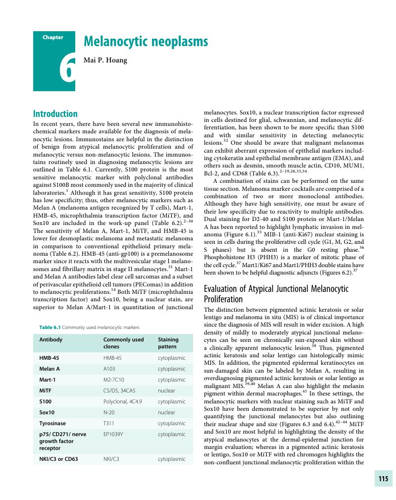
- Chapter 6 Melanocytic neoplasms
- Chapter 7 Soft Tissue Neoplasms
- Chapter 8 Miscellaneous Tumors
- Chapter 9 Detection of Genetic Syndromes
- Chapter 10 Immunobullous Disorders
- Chapter 11 Cutaneous Infections
- Chapter 12 Therapeutic and Prognostic Applications
- Index
- References
Chapter 6 - Melanocytic neoplasms
Published online by Cambridge University Press: 04 November 2017
By
Edited by

Book contents
- Immunohistochemistry in Diagnostic Dermatopathology
- Immunohistochemistry in Diagnostic Dermatopathology
- Copyright page
- Dedication
- Contents
- Contributors
- Preface and Acknowledgments
- Chapter 1 Introduction to Immunohistochemistry
- Chapter 2 Epithelial or Squamous Neoplasms
- Chapter 3 Neoplasms of Cutaneous Appendages
- Chapter 4 Inflammatory Dermatoses Mimicking Lymphomas
- Chapter 5 Cutaneous Lymphoid Neoplasms
- Chapter 6 Melanocytic neoplasms
- Chapter 7 Soft Tissue Neoplasms
- Chapter 8 Miscellaneous Tumors
- Chapter 9 Detection of Genetic Syndromes
- Chapter 10 Immunobullous Disorders
- Chapter 11 Cutaneous Infections
- Chapter 12 Therapeutic and Prognostic Applications
- Index
- References
Summary
A summary is not available for this content so a preview has been provided. Please use the Get access link above for information on how to access this content.
- Type
- Chapter
- Information
- Immunohistochemistry in Diagnostic Dermatopathology , pp. 115 - 137Publisher: Cambridge University PressPrint publication year: 2017
References
Timar, J, Udvarhelyi, N, Banfalvi, T, Gilde, K, Orosz, Z. Accuracy of the determination of S100B protein expression in malignant melanoma using polyclonal or monoclonal antibodies. Histopathology 2004;44(2):180–84.Google Scholar
Anstey, A, Cerio, R, Ramnarain, N, et al. Desmoplastic malignant melanoma. An immunocytochemical study of 25 cases. Am J Dermatopathol 1994;16(1):14–22.CrossRefGoogle ScholarPubMed
Aung, PP, Sarlomo-Rikala, M, Lasota, J, et al. KBA62 and PNL2: 2 new melanoma markers-immunohistochemical analysis of 1563 tumors including metastatic, desmoplastic, and mucosal melanomas and their mimics. Am J Surg Pathol 2012;36(2):265–72.CrossRefGoogle ScholarPubMed
Busam, KJ, Iversen, K, Coplan, KC, Jungbluth, AA. Analysis of microphthalmia transcription factor expression in normal tissues and tumors, and comparison of its expression with S-100 protein, gp100, and tyrosinase in desmoplastic melanoma. Am J Surg Pathol 2001;25(2):197–204.CrossRefGoogle Scholar
Cohen-Knafo, E, al Saati, T, Aziza, J, et al. Production and characterization of an antimelanoma monoclonal antibody KBA.62 using a new melanoma cell line reactive on paraffin wax embedded sections. J Clin Pathol 1995;48(9):826–31.Google Scholar
Ferenczi, K, Lastra, RR, Farkas, T, et al. MUM-1 expression differentiates tumors in the PEComa family from clear cell sarcoma and melanoma. Int J Surg Pathol 2012;20(1):29–36.Google Scholar
Koch, MB, Shih, IM, Weiss, SW, Folpe, AL. Microphthalmia transcription factor and melnoma cell adhesion molecule expression distinguish desmoplastic/spindle cell melanoma from morphologic mimics. Am J Surg Pathol 2001;25(1):58–64.Google Scholar
Mangini, J, Li, N, Bhawan, J. Immunohistochemical markers of melanocytic lesions: A review of their diagnostic usefulness. Am J Dermatopathol 2002;24(3):270–81.Google Scholar
Miettinen, M, Fernandez, M, Franssila, K, et al. Microphthalmia transcription factor in the immunohistochemical diagnosis of metastatic melanoma: Comparison with four other melanoma markers. Am J Surg Pathol 2001;25(2):205–11.CrossRefGoogle ScholarPubMed
Nonaka, D, Chiriboga, L, Rubin, BP. Differential expression of S100 protein subtypes in malignant melanoma, and benign and malignant peripheral nerve sheath tumors. J Cutan Pathol 2008;35(11):1014–19.CrossRefGoogle ScholarPubMed
Orchard, GE. Comparison of immunohistochemical labelling of melanocyte differentiation antibodies melan-A, tyrosinase and HMB45 with NKIC3 and S100 protein in the evaluation of benign and malignant melanoma. Histochem J 2000;32(8):475–81.CrossRefGoogle Scholar
Pages, C, Rochaix, P, al Saati, T, et al. KBA.62: A useful marker for primary and metastatic melanomas. Hum Pathol 2008;39(8):1136–42.CrossRefGoogle ScholarPubMed
Plaza, JA, Suster, D, Perez-Montiel, D. Expression of immunohistochemical markers in primary and metastatic malignant melanoma: A comparative study in 70 patients using a tissue microarray technique. Appl Immunohistochem Mol Morphol 2007;15(4):421–25.Google Scholar
Rochaix, P, Lacroiz-Triki, M, Lamant, L, et al. PNL2, a new monoclonal antibody directed against a fixative-resistant melanocyte antigen. Mod Pathol 2003;16(5):481–90.Google Scholar
Sundram, U, Harvell, JD, Rouse, RV, Natkunam, Y. Expression of the B-cell proliferation marker MUM1 by melanocytic lesions and comparison with S100, gp100 (HMB45), and MelanA. Mod Pathol 2003;16(8):802–10.CrossRefGoogle ScholarPubMed
Shih, IM, Nesbit, M, Herlyn, M, Kurman, RJ. A new Mel-CAM (CD146)-specific monoclonal antibody, MN-4, on paraffin-embedded tissue. Mod Pathol 1998;11(11):1098–106.Google ScholarPubMed
Shin, J, Vincent, JG, Cuda, JD, et al. Sox10 is expressed in primary melanocytic neoplasms of various histologies but not in fibrohistiocytic proliferations and histiocytoses. J Am Acad Dermatol 2012;67(4):717–26.CrossRefGoogle Scholar
Wick, MR, Swanson, PE, Rocamora, A. Recognition of malignant melanoma by monoclonal antibody HMB-45: An immunohistochemical study of 200 paraffin-embedded cutaneous tumors. J Cutan Pathol 1988;15(4):201–7.Google Scholar
Zubovits, J, Buzney, E, Yu, L, Duncan, LM. HMB-45, S-100, NK1/C3, and MART-1 in metastatic melanoma. Hum Pathol 2004;35(2):217–23.Google Scholar
Skelton, HG 3rd, Smith, KJ, Barrett, TL, Lupton, GP, Graham, JH. HMB-45 staining in benign and malignant melanocytic lesions. A reflection of cellular activation. Am J Dermatopathol 1991;13(6):543–50.Google Scholar
Romano, RC, Carter, JM, Folpe, AL. Aberrant intermediate filament and synaptophysin expression is a frequent event in malignant melanoma: An immunohistochemical study of 73 cases. Mod Pathol 2015;28(8):1033–42.CrossRefGoogle ScholarPubMed
Ben-Izhak, O, Stark, P, Levy, R, et al. Epithelial markers in malignant melanoma. A study of primary lesions and their metastases. Am J Dermatopathol 1994;16(3):241–46.Google Scholar
Sanders, DS, Evans, AT, Allen, CA, et al. Classification of CEA-related positivity in primary and metastatic malignant melanoma. J Pathol 1994;172(4):343–48.CrossRefGoogle ScholarPubMed
Carlson, JA, Dickersin, GR, Sober, AJ, Barnhill, RL. Desmoplastic neurotrophic melanoma. Cancer 1995;75(2):478–94.Google Scholar
Bishop, PW, Menasce, LP, Yates, AJ, Win, NA, Banerjee, SS. An immunophenotypic survey of malignant melanomas. Histopathology 1993;23(2):159–66.Google Scholar
Shah, IA, Gani, OS, Wheler, L. Comparative immunoreactivity of CD68 and HMB-45 in malignant melanoma, neural tumors and nevi. Pathol Res Pract 1997;193(7):497–502.Google Scholar
Donato, R, Sorci, G, Riuzzi, F, et al. S100B’s double life: Intracellular regulator and extracellular signal. Biochim Biophys Acta 2009;1793(6):1008–22.Google Scholar
Ohsie, SJ, Sarantopoulos, GP, Cochran, AJ, et al. Immunohistochemical characteristics of melanoma. J Cutan Pathol 2008;35(5):433–44.CrossRefGoogle ScholarPubMed
Thum, C, Hollowood, K, Birch, J, Goodlad, JR, Brenn, T. Aberrant Melan-A expression in atypical fibroxanthoma and undifferentiated pleomorphic sarcoma of the skin. J Cutan Pathol 2011;38(12):954–60.Google Scholar
Miettinen, M, McCue, PA, Sariomo-Rikala, M, et al. Sox-10 – A marker for not only schwannian and melanocytic neoplasms but also myoepithelial cell tumors of soft tissue: A systematic analysis of 5134 tumors. Am J Surg Pathol 2015;39(6): 826–35.Google Scholar
Lee, ZH, Hou, L, Moellmann, G, et al. Characterization and subcellular localization of human Pmel 17/silver, a 110-kDa (pre)melanosomal membrane protein associated with 5,6-dihydroxyindole-2-carboxylic acid (DHICA) converting activity. J Invest Dermatol 1996;106(4):605–10.CrossRefGoogle ScholarPubMed
Nonaka, D, Chiriboga, L, Rubin, BP. Sox10: A pan-schwannian and melanocytic marker. Am J Surg Pathol 2008;32(9):1291–98.CrossRefGoogle ScholarPubMed
Busam, KJ, Kucukgol, D, Sato, E, et al. Immunohistochemical analysis of novel monoclonal antibody PNL2 and comparison with other melanocyte differentiation markers. Am J Surg Pathol 2005;29(3):400–6.Google Scholar
Banerjee, SS, Harris, M. Morphological and immunophenotypic variations in malignant melanoma. Histopathology 2000;36(5):387–402.Google Scholar
Petitt, M, Allison, A, Shimoni, T, et al. Lymphatic invasion detected by D2-40/S100 dual immunohistochemistry does not predict sentinel lymph node status in melanoma. J Am Acad Dermatol 2009;61(5):819–28.Google Scholar
Gerdes, J, Lemke, H, Baisch, H, et al. Cell cycle analysis of a cell proliferation-associated human nuclear antigen defined by the monoclonal antibody Ki-67. J Immunol 1984;133(4):1710–15.CrossRefGoogle ScholarPubMed
Ikenberg, K, Pfaltz, M, Rakozy, C, Kempf, W. Immunohistochemical dual staining as an adjunct in assessment of mitotic activity in melanoma. J Cutan Pathol 2012;39(3):324–30.CrossRefGoogle ScholarPubMed
Hendi, A, Wada, DA, Jacobs, MA, Crook, JE, Korteum, KR, et al. Melanocytes in nonlesional sun-exposed skin: A multicenter comparative study. J Am Acad Dermatol 2011;65(6):1186–93.CrossRefGoogle ScholarPubMed
Beltraminelli, H, Shabrawi-Caelen, LE, Kerl, H, Cerroni, L. Melan-a-positive “pseudomelanocytic nests”: A pitfall in the histopathologic and immunohistochemical diagnosis of pigmented lesions on sun-damage skin. Am J Dermatopathol 2009;31(3):305–8.CrossRefGoogle Scholar
El Shabrawi-Caelen, L, Kerl, H, Cerroni, L. Melan-A: Not a helpful marker in distinction between melanoma in situ on sun-damaged skin and pigmented actinic keratosis. Am J Dermatopathol. 2004;26(5):364–6.Google Scholar
Yan, S, Brennick, JB. False-positive rate of the immunoperoxidase stains for MART1/MelanA in lymph nodes. Am J Surg Pathol 2004;28(5):596–600.CrossRefGoogle ScholarPubMed
Kim, J, Taube, JM, McCalmont, TH, Glusac, EJ. Quantitative comparison of MiTF, Melan-A, HMB-45 and Mel-5 in solar lentigines and melanoma in situ. J Cutan Pathol 2011;38(10):775–79.Google ScholarPubMed
Glass, LF, Raziano, RM, Clark, GS, et al. Rapid frozen section immunostaining of melanocytes by microphthalmia-associated transcription factor. Am J Dermatopathol 2010;32(4):319–25.Google Scholar
Buonaccorsi, JN, Prieto, VG, Torres-Cabala, C, Suster, S, Plaza, JA. Diagnostic utility and comparative immunohistochemical analysis of MiTF and Sox10 to distinguish melanoma in situ and actinic keratosis: A clinicopathological and immunohistochemical study of 70 cases. Am J Dermatopathol 2014;36(2):124–30.Google Scholar
Ramos-Herberth, FI, Karamchandani, J, Kim, J, Dadras, SS. Sox10 immunostaining distinguishes desmoplastic melanoma from excision scar. J Cutan Pathol 2010;37(9):944–52.Google Scholar
Harvell, JD, Bastian, BC, LeBoit, PE. Persistent (recurrent) Spitz nevi: A histopathologic, immunohistochemical, and molecular pathologic study of 22 cases. Am J Surg Pathol 2002;26(5):654–61.Google Scholar
Wood, WS, Tron, VA. Analysis of HMB-45 immunoreactivity in common and cellular blue nevi. J Cutan Pathol 1991;18(4):261–63.Google Scholar
McNutt, NS, Urmacher, C, Hakimian, J, Hoss, DM, Lugo, J. Nevoid malignant melanoma: Morphologic patterns and immunohistochemical reactivity. J Cutan Pathol 1995;22(6):502–17.CrossRefGoogle ScholarPubMed
Prieto, VG, Shea, CR. Use of immunohistochemistry in melanocytic lesions. J Cutan Pathol 2008;35(Suppl 2):1–10.Google Scholar
Rothberg, BEG, Moeder, CB, Kluger, H, et al. Nuclear to non-nuclear Pmel17/gp100 expression (HMB45 staining) as a discriminator between benign and malignant melanocytic lesions. Mod Pathol 2008;21(9):1121–29.Google Scholar
Puri, PK, Elston, CA, Tyler, WB, Ferringer, TC, Elston, DM. The staining pattern of pigmented spindle cell nevi with S100A6 protein. J Cutan Pathol 2011;38(1):14–17.CrossRefGoogle ScholarPubMed
Nielsen, PS, Riber-Hansen, R, Raundahl, J, Steiniche, T. Automated quantification of MART1-verified Ki67 indices by digital image analysis in melanocytic lesions. Arch Pathol Lab Med 2012;136(6):627–34.Google Scholar
Kamino, H, Tam, S, Tapia, B, Toussaint, S. The use of elastin immunostain improves the evaluation of melanomas associated with nevi. J Cutan Pathol 2009;36(8):845–52.Google Scholar
Bergman, R, Malkin, L, Sabo, E, Kerner, H. MIB-1 monoclonal antibody to determine proliferative activity of Ki-67 antigen as an adjunct to the histopathologic differential diagnosis of Spitz nevi. J Am Acad Dermatol 2001;44(3):500–4.Google Scholar
Kanter-Lewensohn, L, Hedblad, MA, Wejde, J, Larsson, O. Immunohistochemical markers for distinguishing Spitz nevi from malignant melanomas. Mod Pathol 1997;10(9):917–20.Google ScholarPubMed
Kapur, P, Selim, MA, Roy, LC, et al. Spitz nevi and atypical Spitz nevi/tumors: A histologic and immunohistochemical analysis. Mod Pathol 2005;18(2):197–204.Google Scholar
Gurley, LR, D’Anna, JA, Barham, SS, Deaven, LL, Tobey, RA. Histone phosphorylation and chromatin structure during mitosis in Chinese hamster cells. Eur J Biochem 1978;84(1):1–15.CrossRefGoogle ScholarPubMed
Tapia, C, Kutzner, H, Mentzel, T, et al. Two mitosis-specific antibodies, MPM-2 and phospho-histone H3 (Ser28), allow rapid and precise determination of mitotic activity. Am J Surg Pathol 2006;30(1):83–89.CrossRefGoogle ScholarPubMed
Nasr, MR, El-Zammar, O. Comparison of PHH3, Ki-67, and surviving immunoreactivity in benign and malignant melanocytic lesions. Am J Dermatopathol 2008;30(2):117–22.CrossRefGoogle Scholar
Phadke, PA, Rakheja, D, Le, LP, et al. Proliferative nodules arising within congenital melanocytic nevi: A histologic, immunohistochemical, and molecular analyses of 43 cases. Am J Surg Pathol 2011;35(5):656–69.Google Scholar
Herron, MD, Vanderhooft, SL, Smock, K, et al. Proliferative nodules in congenital melanocytic nevi: A clinicopathologic and immunohistochemical analysis. Am J Surg Pathol 2004;28(8):1017–25.Google Scholar
Ludgate, MW, Fullen, DR, Lee, J, et al. The atypical Spitz tumor of uncertain biologic potential: A series of 67 patients from a single institution. Cancer 2009;115(3):631–41.Google Scholar
Massi, D, Tomasini, C, Senetta, R, et al. Atypical Spitz tumors in patients younger than 18 years. J Am Acad Dermatol 2015;72(1):37–46.Google Scholar
Bergman, R, Dromi, R, Trau, H, Cohen, I, Lichtig, C. The pattern of HMB-45 antibody staining in compound Spitz nevi. Am J Dermatopathol 1995;17(6):542–46.Google Scholar
Puri, PK, Ferringer, TC, Tyler, WB, et al. Statistical analysis of the concordance of immunohistochemical stains with the final diagnosis of spitzoid neoplasms. Am J Dermatopathol 2011;33(1):72–77.Google Scholar
Plaza, JA, De Stafano, D, Suster, S, et al. Intradermal Spitz nevi: A rare subtype of Spitz nevi analyzed in a clinicopathologic study of 74 cases. Am J Dermatopathol 2014;36(4):283–94.CrossRefGoogle Scholar
Vollmer, RT. Use of Bayes rule and MIB-1 proliferation index to discriminate Spitz nevus from malignant melanoma. Am J Clin Pathol 2004;122(4):499–505.Google Scholar
George, E, Polissar, NL, Wick, M. Immunohistochemical evaluation of p16INK4A, E-cadherin, and cyclin D1 expression in melanoma and Spitz tumors. Am J Clin Pathol 2010;133(3):370–79.Google Scholar
Ribe, A, McNutt, NS. S100A6 protein expression is different in Spitz nevi and melanomas. Mod Pathol 2003;16(5):505–11.CrossRefGoogle ScholarPubMed
Gerami, P, Scolyer, RA, Xu, X, et al. Risk assessment for atypical spitzoid melanocytic neoplasms using FISH to identify chromosomal copy number aberrations. Am J Surg Pathol 2013;37(5):676–84.Google Scholar
Shen, L, Cooper, C, Bajaj, S, et al. Atypical spitz tumors with 6q23 deletions: A clinical, histological, and molecular study. Am J Dermatopathol 2013;35(8):804–12.Google Scholar
Wiesner, T, Murali, R, Fried, I, et al. A distinct subset of atypical Spitz tumors is characterized by BRAF mutation and loss of BAP1 expression. Am J Surg Pathol 2012;36(6):818–30.Google Scholar
Yazdan, P, Cooper, C, Sholl, LM, et al. Comparative analysis of atypical Spitz tumors with heterozygous versus homozygous 9p21 deletions for clinical outcomes, histomorphology, BRAF mutation, and p16 expression. Am J Surg Pathol 2014;38(5):638–45.CrossRefGoogle ScholarPubMed
Van Dijk, MCRF, Bernsen, MR, Ruiter, DJ. Analysis of mutations in B-RAF, N-RAS, and H-RAS genes in the differential diagnosis of Spitz nevus and Spitzoid melanoma. Am J Surg Pathol 2005;29(9):1145–51.Google Scholar
Cooper, C, Arva, NC, Lee, C, et al. A clinical, histopathologic, and outcome study of melanonychia striata in childhood. J Am Acad Dermatol 2015;72(5):773–79.Google Scholar
Tan, KB, Moncrieff, M, Thompson, JF, et al. Subungual melanoma: A study of 124 cases highlighting features of early lesions, potential pitfalls in diagnosis, and guidelines for histologic reporting, Am J Surg Pathol 2007;31(12):1902–12.CrossRefGoogle ScholarPubMed
Theunis, A, Richert, B, Sass, U, et al. Immunohistochemical study of 40 cases of longitudinal melanonychia. Am J Dermatopathol 2011;33(1):27–34.Google Scholar
Ridolfi, RL, Rosen, PP, Thaler, H. Nevus cell aggregates associated with lymph nodes: Estimated frequency and clinical significance. Cancer 1977;39(1):164–71.Google Scholar
Carson, KF, Wen, DR, Li, PX, et al. Nodal nevi and cutaneous melanoma. Am J Surg Pathol 1996;20(7):834–40.Google Scholar
Yan, S, Brennick, JB. False-positive rate of the immunoperoxidase stains for MART1/MelanA in lymph nodes. Am J Surg Pathol 2004;28(5):596–600.CrossRefGoogle ScholarPubMed
Biddle, DA, Evans, HL, Kemp, BL, et al. Intraparenchymal nevus cell aggregates in lymph nodes: A possible diagnostic pitfalls with malignant melanoma and carcinoma. Am J Surg Pathol 2003;27(5):673–81.Google Scholar
Holt, JB, Sangueza, OP, Levine, EA, et al. Nodal melanocytic nevi in sentinel lymph nodes. Correlation with melanoma-associated cutaneous nevi. Am J Clin Pathol 2004;121(1):58–63.CrossRefGoogle ScholarPubMed
Lohmann, CM, Iversen, K, Jungbluth, AA, Berwick, M, Busam, KJ. Expression of melanocyte differentiation antigens and ki-67 in noval nevi and comparison of Ki-67 expression with metastatic melanoma. Am J Surg Pathol 2002;26(10):1351–57.Google Scholar
Mihic-Probst, D, Saremaslani, P, Komminoth, P, Heitz, PU. Immunostaining for the tumour suppressor gene p16 product is a useful marker to differentiate melanoma metastasis from lymph-node nevus. Virchows Arch 2003;443(6):745–51.CrossRefGoogle ScholarPubMed
Blochin, E, Nonaka, D. Diagnostic value of Sox10 immunohistochemical staining for the detection of metastatic melanoma in sentinel lymph nodes. Histopathology 2009;55(5):626–28.Google Scholar
Piana, S, Tagliavini, E, Tagazzi, M, et al. Lymph node melanocytic nevi: Pathogenesis and differential diagnoses, with special reference to p16 reactivity. Pathol Res Pract 2015;211(5):381–88.Google Scholar
Kanner, WA, Barry, CI, Smart, CN, et al. Reticulin and NM23 staining in the interpretation of lymph node nevus rests. Am J Dermatopathol 2013;35(4)452–57.Google Scholar
Mentrikoshi, MJ, Ma, L, Pryor, JG, et al. Diagnostic utility of IMP3 in segregating metastatic melanoma from benign nevi in lymph nodes. Mod Pathol 2009;22(12):1582–87.Google Scholar
Chen, PL, Chen, WS, Li, J, Lind, AC, Lu, D. Diagnostic utility of neural stem and progenitor cell markers nestin and SOX2 in distinguishing nodal melanocytic nevi from metastatic melanomas. Mod Pathol 2013;26(1):44–53.Google Scholar
Lee, JJ, Granter, SR, Laga, AC, et al. 5-hydroxymethylcytosine expression in metastatic melanoma versus nodal nevus in sentinel lymph node biopsies. Mod Pathol 2015;28(2):218–29.Google Scholar
Palla, B, Su, A, Binder, S, Dry, S. Sox10 expression distinguishes desmoplastic melanoma from its histologic mimics. Am J Dermatopathol 2013;35(5):576–81.CrossRefGoogle ScholarPubMed
Kucher, C, Zhang, PJ, Pasha, T, et al. Expression of Melan-A and Ki-67 in desmoplastic melanoma and desmoplastic nevi. Am J Dermatopathol 2004;26(6):452–57.Google Scholar
Lazova, R, Tantcheva-Poor, I, Sigal, AC. P75 nerve growth factor receptor staining is superior to S100 in identifying spindle cell and desmoplastic melanoma. J Am Acad Dermatol 2010;63(5):852–58.Google Scholar
Plaza, JA, Bonneau, P, Prieto, V, et al. Desmoplastic melanoma: An updated immunohistochemical analysis of 40 cases with a proposal for an additional panel of stains for diagnosis. J Cutan Pathol 2016;43(4):313–23.CrossRefGoogle ScholarPubMed
Chorny, JA, Barr, RJ. S100-positive spindle cells in scars: A diagnostic pitfall in the re-excision of desmoplastic melanoma. Am J Dermatopathol 2002;24(4):309–12.Google Scholar
Otaibi, S, Jukic, DM, Drogowski, L, Bhawan, J, Radfar, A. NGFR (p75) expression is cutaneous scars; further evidence for a potential pitfall in evaluation of reexcision scars of cutaneous neoplasms, in particular desmoplastic melanoma. Am J Dermatopathol 2011;33(1):65–71.Google Scholar
Ramos-Herberth, FI, Karamchandani, J, Kim, J, Dadras, SS. Sox10 immunostaining distinguishes desmoplastic melanoma from excision scar. J Cutan Pathol 2010;37(9):944–52.Google Scholar
Meis-Kindblom, JM. Clear cell sarcoma of tendons and aponeuroses: A historical perspective and tribute to the man behind the entity. Adv Anat Pathol 2006;13(6):186–292.CrossRefGoogle Scholar
Bianchi, G, Charoenlap, C, Cocchi, S, et al. Clear cell sarcoma of soft tissue: A retrospective review and analysis of 31 cases treated at Istituto Ortopedico Rizzoli. Eur J Surg Oncol 2014;40(5):505–10.Google Scholar
Hisaoka, M, Ishida, T, Kuo, TT, et al. Clear cell sarcoma of soft tissue: A clinicopathologic, immunohistochemical, and molecular analysis of 33 cases. Am J Surg Pathol 2008;32(3):452–60.Google Scholar
Coindre, JM, Hostein, I, Terrier, P, et al. Diagnosis of clear cell sarcoma by real-time reverse transcriptase-polymerase chain reaction analysis of paraffin embedded tissues: Clinicopathologic and molecular analysis of 44 patients from the French sarcoma group. Cancer 2006;107(1):1055–64.Google Scholar
Granter, SR, Weilbaecher, KN, Quigley, C, Fletcher, CD, Fisher, DE. Clear cell sarcoma shows immunoreactivity for microphthalmia transcription factor: Further evidence for melanocytic differentiation. Mod Pathol 2001;14(1):6–9.Google Scholar
Aung, PP, Sarlomo-Rikala, M, Lasota, J, et al. KBA62 and PNL2: 2 new melanoma markers- immunohistochemical analysis of 1563 tumors including metastatic, desmoplastic, and mucosal melanomas and their mimics. Am J Surg Pathol 2012;36(2):265–72.Google Scholar
Hantschke, M, Mentzel, T, Rutten, A, et al. Cutaneous clear cell sarcoma: A clinicopathologic, immunohistochemical, and molecular analysis of 12 cases emphasizing its distinction from dermal melanoma. Am J Surg Pathol 2010;34(2):216–22.Google Scholar
Karamchandani, JR, Nielsen, TO, van de Rijn, M, West, RB. Sox10 and S100 in the diagnosis of soft-tissue neoplasms. Appl Immunohistochem Mol Morphol 2012;20(5):445–50.Google Scholar
Wang, WL, Mayordomo, E, Zhang, W, et al. Detection and characterization of EWSR1/ATF1 and EWSR1/CREB1 chimeric transcripts in clear cell sarcoma (melanoma of soft parts). Mod Pathol 2009;22(9):1201–9.Google Scholar
Yang, L, Chen, Y, Cui, T, et al. Identification of biomarkers to distinguish clear cell sarcoma from malignant melanoma. Hum Pathol 2012;43(9):1463–70.Google Scholar
Charli-Joseph, Y, Saggini, A, Vemula, S, et al. Primary cutaneous perivascular epithelioid cell tumor: A clinicopathological and molecular reappraisal. J Am Acad Dermatol 2014;71(6):1127–36.Google Scholar
Liegl, B, Hornick, JL, Fletcher, CDM. Primary cutaneous PEComa: Distinctive clear cell lesions of skin. Am J Surg Pathol 2008;32(4):608–14.Google Scholar
Llamas-Velasco, M, Mentzel, T, Requena, L, et al. Cutaneous PEComa does not harbour TFE3 gene fusions: Immunohistochemical and molecular study of 17 cases. Histopathology 2013;63(1):122–29.Google Scholar
Mentzel, T, Reisshauer, S, Rutten, A, Hantschke, M, Soares de Almeida, LM, Kutzner, H. Cutaneous clear cell myomelanocytic tumour: A new member of the growing family of perivascular peithelioid cell tumours (PEComas). Clinicopathological and immunohistochemical analysis of seven cases. Histopathology 2005;46(5):498–504.CrossRefGoogle ScholarPubMed
Greveling, K, Winnepennickx, VJ, Nagtzaam, IF, et al. Malignant perivascular epithelioid tumor: A case report of a cutaneous tumor on the cheek of a male patient. J Am Acad Dermatol 2013;69(5):e262–64.CrossRefGoogle ScholarPubMed
Calder, KB, Schlauder, S, Morgan, MB. Malignant perivascular epithelioid cell tumor (“PEComa”): A case report and literature review of cutaneous/subcutaneous presentations. J Cutan Pathol 2008;35(5):499–503.Google Scholar
Martignoni, G, Gobbo, S, Camparo, P, et al. Differential expression of cathepsin K in neoplasms harboring TFE3 gene fusions. Mod Pathol 2011;24(10):1313–19.Google Scholar
Tallon, B, Beer, TW. MiTF positivity in atypical fibroxanthoma: A diagnostic pitfall. Am J Dermatopathol 2014;36(11):888–91.Google Scholar
Suarez-Vilela, D, Izquierdo, FM, Escobar-Stein, J, Mendez-Alvarez, JR. Atypical fibroxanthoma with T-cytotoxic inflammatory infiltrate and aberrant expression of cytokeratin. J Cutan Pathol 2011;38(1):930–32.Google Scholar
Uquen, A, Sassolas, B, Mondine, P, et al. NRASQ61 R and BRAFV600E mutation-specific immunohistochemistry is a helpful tool to diagnose metastatic undifferentiated/dedifferentiated melanomas. Am J Surg Pathol 2016;40(7):1004–5.Google Scholar
Henderson, SA, Torres-Cabala, CA, Curry, JL, et al. p40 is more specific than p63 for the distinction of atypical fibroxanthoma from other cutaneous spindle cell malignancies. Am J Surg Pathol 2014;38(8):1102–10.CrossRefGoogle ScholarPubMed
Kao, GF, Helwig, EB, Graham, JH. Balloon cell malignant melanoma of the skin. A clinicopathologic study of 34 cases with histochemical, immunohistochemical, and ultrastructural observations. Cancer 1992;69(12):2942–52.Google Scholar
Plaza, JA, Torres-Cabala, C Evans, H, et al. Cutaneous metastases of malignant melanoma: A clinicopathologic study of 192 cases with emphasis on the morphologic spectrum. Am J Dermatopathol 2010;32(2):129–36.CrossRefGoogle ScholarPubMed
- 1
- Cited by