
- Publisher:
- Cambridge University Press
- Online publication date:
- May 2016
- Print publication year:
- 2016
- Online ISBN:
- 9781316182123
- Subjects:
- Medical Imaging, Emergency Medicine, Medicine
Our systems are now restored following recent technical disruption, and we’re working hard to catch up on publishing. We apologise for the inconvenience caused. Find out more: https://www.cambridge.org/universitypress/about-us/news-and-blogs/cambridge-university-press-publishing-update-following-technical-disruption
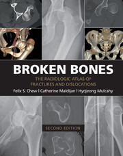
Broken Bones contains 434 individual cases and 1,101 radiologic images illustrating the typical and less typical appearances of fractures and dislocations throughout the body. The first chapter describes fractures and dislocations of the fingers, starting with fractures of the phalangeal tufts and progressing through the distal, middle, and proximal phalanges and the DIP and PIP joints. Subsequent chapters cover the metacarpals, the carpal bones, the radius and ulna, the elbow and upper arm, and the shoulder and thoracic cage. The cervical spine and the thoracic and lumbosacral spine are covered in separate chapters, followed by the pelvis, the femur, the knee and lower leg, the ankle, the tarsal bones, and the metatarsals and toes. The final three chapters cover the face, fractures and dislocations in children, and fractures and dislocations caused by bullets and nonmilitary blasts.
 Loading metrics...
* Views captured on Cambridge Core between #date#. This data will be updated every 24 hours.
Usage data cannot currently be displayed.